File:Bailey317-319.jpg
From Embryology
Size of this preview: 368 × 599 pixels. Other resolution: 663 × 1,080 pixels.
Original file (663 × 1,080 pixels, file size: 99 KB, MIME type: image/jpeg)
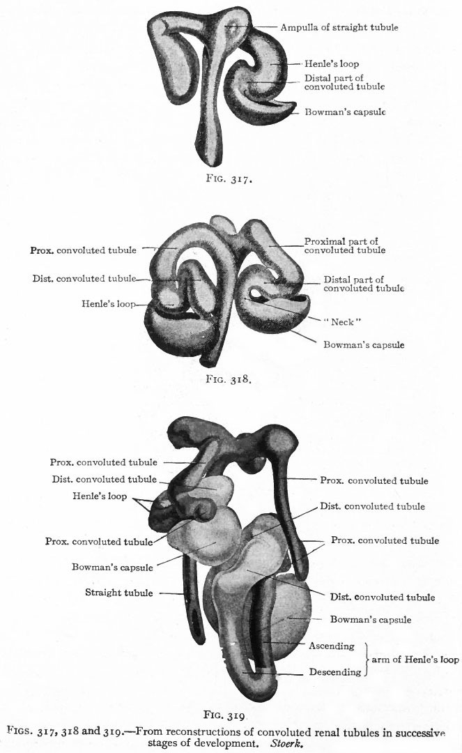
Figs. 317, 318 and 319. From reconstructions of convoluted renal tubules in successive stages of development
Stoerk.
The Convoluted Renal Tubules and Glomeruli.
- As stated above, the metanephric blastema or nephrogenic tissue surrounds the renal pelvis and the straight tubules.
- It represents a condensation of the mesenchyme and is destined to give rise to the convoluted tubules and glomeruli.
- The cells of the blastema in the region of the ampullae of the terminal straight tubules acquire an epithelial character and become arranged in solid masses (Fig. 315).
- Each mass unites with an ampulla and acquires a lumen, which becomes continuous with the lumen of the straight tubule, then elongates and forms an S-shaped structure (Figs. 316 and 317).
- The loop of the S nearer the straight tubules elongates still more and grows toward the pelvis, parallel with the straight tubules, to form Henle's loop.
- The part between Henle's loop and the straight tubule elongates and becomes convoluted to form the proximal part of a convoluted renal tubule (second convoluted tubule) .
- The part between the distal end and Henle's loop elongates and becomes convoluted to form the distal part of a convoluted renal tubule (first convoluted tubule) (Figs. 318 and 319).
- Link: Figure in Text
- Text-Book of Embryology: Germ cells | Maturation | Fertilization | Amphioxus | Frog | Chick | Mammalian | External body form | Connective tissues and skeletal | Vascular | Muscular | Alimentary tube and organs | Respiratory | Coelom, Diaphragm and Mesenteries | Urogenital | Integumentary | Nervous System | Special Sense | Foetal Membranes | Teratogenesis | Gallery of All Figures
| Historic Disclaimer - information about historic embryology pages |
|---|
 |
Reference
Bailey FR. and Miller AM. Text-Book of Embryology (1921) New York: William Wood and Co.
Cite this page: Hill, M.A. (2024, April 24) Embryology Bailey317-319.jpg. Retrieved from https://embryology.med.unsw.edu.au/embryology/index.php/File:Bailey317-319.jpg
- © Dr Mark Hill 2024, UNSW Embryology ISBN: 978 0 7334 2609 4 - UNSW CRICOS Provider Code No. 00098G
File history
Click on a date/time to view the file as it appeared at that time.
| Date/Time | Thumbnail | Dimensions | User | Comment | |
|---|---|---|---|---|---|
| current | 11:30, 25 January 2011 | | 663 × 1,080 (99 KB) | S8600021 (talk | contribs) | {{Template:Bailey 1921 Figures}} Category:Human Category:Renal Category:Bladder |
You cannot overwrite this file.
File usage
The following 2 pages use this file: