File:Bailey312.jpg
Original file (823 × 839 pixels, file size: 82 KB, MIME type: image/jpeg)
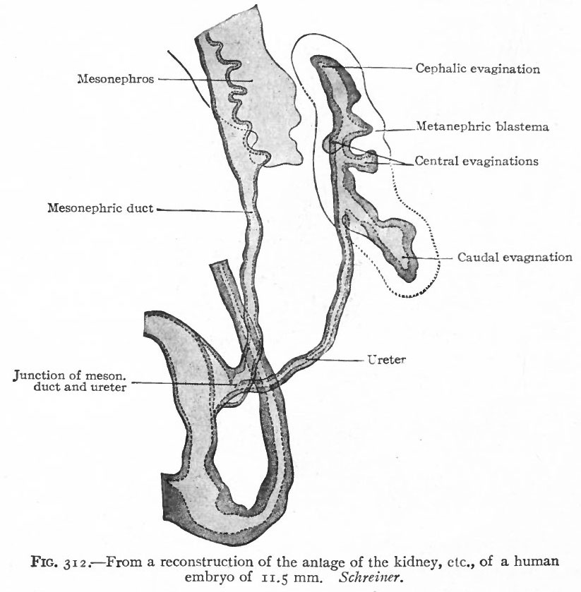
Fig. 312. From a reconstruction of the anlage of the kidney of a human embryo of 11.5 mm
Schreiner.
When the dilated end reaches the ventral side of the vertebral column it turns and grows cranially between the latter and the mesonephros. The stalk (or ureter) elongates accordingly (Fig. 312).
About the fifth week, four evaginations from the primitive renal pelvis appear one cephalic, one caudal and two central (Figs. 312 and 314) . These may be considered as straight renal tubules of the first order. The distal end of each then enlarges to form a sort of ampulla, and from each ampulla two other evaginations develop, forming tubules of the second order. From the ampulla of each secondary tubule two tertiary tubules grow out; and this process continues in a similar manner until twelve or thirteen divisions occur, the final divisions occurring during the fifth month.
- Link: Figure in Text
- Text-Book of Embryology: Germ cells | Maturation | Fertilization | Amphioxus | Frog | Chick | Mammalian | External body form | Connective tissues and skeletal | Vascular | Muscular | Alimentary tube and organs | Respiratory | Coelom, Diaphragm and Mesenteries | Urogenital | Integumentary | Nervous System | Special Sense | Foetal Membranes | Teratogenesis | Gallery of All Figures
| Historic Disclaimer - information about historic embryology pages |
|---|
 |
Reference
Bailey FR. and Miller AM. Text-Book of Embryology (1921) New York: William Wood and Co.
Cite this page: Hill, M.A. (2024, April 24) Embryology Bailey312.jpg. Retrieved from https://embryology.med.unsw.edu.au/embryology/index.php/File:Bailey312.jpg
- © Dr Mark Hill 2024, UNSW Embryology ISBN: 978 0 7334 2609 4 - UNSW CRICOS Provider Code No. 00098G
File history
Click on a date/time to view the file as it appeared at that time.
| Date/Time | Thumbnail | Dimensions | User | Comment | |
|---|---|---|---|---|---|
| current | 11:16, 25 January 2011 | | 823 × 839 (82 KB) | S8600021 (talk | contribs) |
You cannot overwrite this file.
File usage
The following 2 pages use this file: