File:Bailey262.jpg
Bailey262.jpg (406 × 482 pixels, file size: 57 KB, MIME type: image/jpeg)
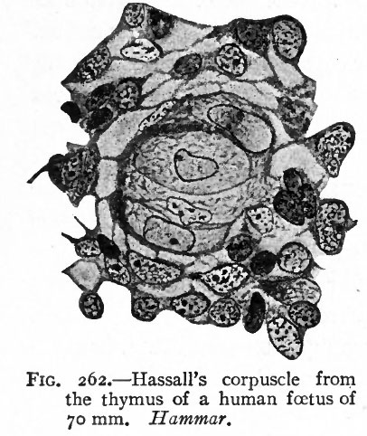
Fig. 262.
The histogenesis of the thymus has been a subject of much study and controversy, not only in regard to its origin, but also in regard to its change from an epithelial to a lymphoid structure and the regressive changes in the latter. It has almost certainly been proven to be of entodermal origin. It is at first an epithelial mass which later becomes broken up into lobules by the ingrowth of connective tissue. In regard to the histological changes which it undergoes, the older views are in general that a " pseudomorphosis " takes place; that is, the epithelial elements are replaced by lymphoid cells which wander in from the neighboring blood vessels, Hassall's corpuscles being remnants of the epithelium. Later other investigators looked upon the changes as a "transformation," asserting that the epithelial cells were transformed into lymphoid cells in situ, and that Hassall's corpuscles were remnants of epithelium and disintegrating blood vessels. Some went even so far as to assert that the thymus was the first place of origin of the leucocytes. More recent researches furnish very strong evidence that no lymphoid cells are derived from the epithelial cells (Maximow), but that the epithelium is transformed into the reticular tissue of the thymus, in which the lymphoid cells undergo mitotic division, Hassall's corpuscles possibly representing compressed parts of the reticulum (Hammar) (Fig. 262).
- Text-Book of Embryology: Germ cells | Maturation | Fertilization | Amphioxus | Frog | Chick | Mammalian | External body form | Connective tissues and skeletal | Vascular | Muscular | Alimentary tube and organs | Respiratory | Coelom, Diaphragm and Mesenteries | Urogenital | Integumentary | Nervous System | Special Sense | Foetal Membranes | Teratogenesis | Gallery of All Figures
| Historic Disclaimer - information about historic embryology pages |
|---|
 |
Reference
Bailey FR. and Miller AM. Text-Book of Embryology (1921) New York: William Wood and Co.
Cite this page: Hill, M.A. (2024, April 25) Embryology Bailey262.jpg. Retrieved from https://embryology.med.unsw.edu.au/embryology/index.php/File:Bailey262.jpg
- © Dr Mark Hill 2024, UNSW Embryology ISBN: 978 0 7334 2609 4 - UNSW CRICOS Provider Code No. 00098G
File history
Click on a date/time to view the file as it appeared at that time.
| Date/Time | Thumbnail | Dimensions | User | Comment | |
|---|---|---|---|---|---|
| current | 21:56, 23 January 2011 | | 406 × 482 (57 KB) | S8600021 (talk | contribs) | {{Template:Bailey 1921 Figures}} Category:Human Category:Gastrointestinal Tract |
You cannot overwrite this file.
File usage
The following 2 pages use this file: