File:Bailey256.jpg

Original file (894 × 545 pixels, file size: 75 KB, MIME type: image/jpeg)
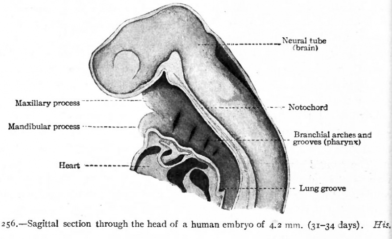
Fig. 256. Sagittal section through the head of a human embryo of 4.2 mm (31-34 days)
The pharynx develops from the cephalic end of the primitive gut. This part of the gut is primarily of uniform diameter, is broadly attached by mesoderm to the dorsal body wall, and ends blindly (Fig. 247). When the branchial arches and grooves develop in this (the cervical) region, they affect the gut as well as the periphery of the body. The arches form ridges on the surface of the body (Fig. 85) and at the same time form ridges on the wall of the gut. The grooves form pockets which alternate with the arches (Fig. 256). The pock in the pharyngeal cavity, or inner branchial grooves, are directed outward toward corresponding outer branchial grooves (Fig. 249). The arches are covered externally with ectoderm, internally with entoderm, and are filled with mesoderm. Between the arches, or in the grooves, the ectoderm and entoden are in contact or nearly so. Thus the pharynx is not surrounded by a coelomic cavity.
- Text-Book of Embryology: Germ cells | Maturation | Fertilization | Amphioxus | Frog | Chick | Mammalian | External body form | Connective tissues and skeletal | Vascular | Muscular | Alimentary tube and organs | Respiratory | Coelom, Diaphragm and Mesenteries | Urogenital | Integumentary | Nervous System | Special Sense | Foetal Membranes | Teratogenesis | Gallery of All Figures
| Historic Disclaimer - information about historic embryology pages |
|---|
 |
Reference
Bailey FR. and Miller AM. Text-Book of Embryology (1921) New York: William Wood and Co.
Cite this page: Hill, M.A. (2024, April 25) Embryology Bailey256.jpg. Retrieved from https://embryology.med.unsw.edu.au/embryology/index.php/File:Bailey256.jpg
- © Dr Mark Hill 2024, UNSW Embryology ISBN: 978 0 7334 2609 4 - UNSW CRICOS Provider Code No. 00098G
File history
Click on a date/time to view the file as it appeared at that time.
| Date/Time | Thumbnail | Dimensions | User | Comment | |
|---|---|---|---|---|---|
| current | 10:36, 21 January 2011 | | 894 × 545 (75 KB) | S8600021 (talk | contribs) | {{Template:Bailey 1921 Figures}} Category:Human Category:Gastrointestinal Tract |
You cannot overwrite this file.
File usage
The following 2 pages use this file: