File:Bailey082.jpg
Original file (879 × 756 pixels, file size: 137 KB, MIME type: image/jpeg)
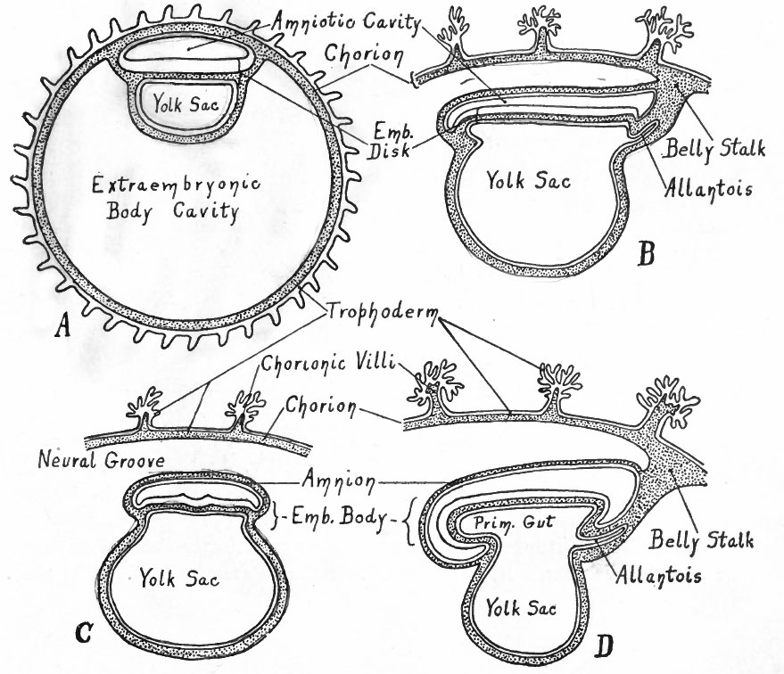
Fig. 82. Diagrams representing stages of development of the human embryo
(to follow Fig. 81).
A - A stage that corresponds approximately to those of Peters' and Bryce-Teacher's embryos (Figs. 74 and 73). Owing to the rapid enlargement of the chorionic vesicle, the extraembryonic body cavity has become much larger than in Fig. 81 , C.
B - A stage (in longitudinal section) corresponding to that of von Spec's embryo (Fig. 77) . Only a part of the chorion is shown; the embryonic disk is slightly constricted from the yolk sac; note the belly stalk, comparing with A .
C - Transverse section, same stage as B.
D - Longitudinal section, stage somewhat later than B. Note the greater degree of constriction between the embryo and yolk sac, and the larger amnion.
The series of diagrams in Figs. 80, 81 and 82 has been constructed to give the student a general idea of the changes that occur in the early stages of human development. It must be recognized, however, that the diagrams represent purely hypothetical stages up to the conditions shown in diagram B in Fig. 81 which corresponds roughly to the Bryce-Teacher embryo (Fig. 73) ; even in this diagram the extent of the mesoderm is much less than in the known human embryo. In Fig. 82 diagram A approximates the Peters embryo (Fig. 74), diagram D the von Spee embryo (Fig. 77). The history of the accessory structures which are shown in part will be considered in the chapter on "Foetal Membranes".
- Text-Book of Embryology: Germ cells | Maturation | Fertilization | Amphioxus | Frog | Chick | Mammalian | External body form | Connective tissues and skeletal | Vascular | Muscular | Alimentary tube and organs | Respiratory | Coelom, Diaphragm and Mesenteries | Urogenital | Integumentary | Nervous System | Special Sense | Foetal Membranes | Teratogenesis | Gallery of All Figures
| Historic Disclaimer - information about historic embryology pages |
|---|
 |
Reference
Bailey FR. and Miller AM. Text-Book of Embryology (1921) New York: William Wood and Co.
Cite this page: Hill, M.A. (2024, April 23) Embryology Bailey082.jpg. Retrieved from https://embryology.med.unsw.edu.au/embryology/index.php/File:Bailey082.jpg
- © Dr Mark Hill 2024, UNSW Embryology ISBN: 978 0 7334 2609 4 - UNSW CRICOS Provider Code No. 00098G
File history
Click on a date/time to view the file as it appeared at that time.
| Date/Time | Thumbnail | Dimensions | User | Comment | |
|---|---|---|---|---|---|
| current | 11:29, 18 January 2011 | | 879 × 756 (137 KB) | S8600021 (talk | contribs) | {{Template:Bailey 1921 Figures}} |
You cannot overwrite this file.
File usage
The following 2 pages use this file: