File:B220849-03.jpg

Original file (1,388 × 1,040 pixels, file size: 346 KB, MIME type: image/jpeg)
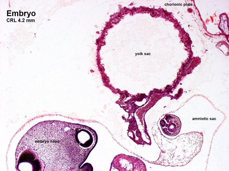
Human Embryo (CRL 4.2 mm)
Sagittal section through embryo week 4 approximately Carnegie stage 11 to 12.
- The section passes through the lateral wall of the embryo head.
- Image shows the extra-embryonic coelomic structures: amniotic sac, yolk sac, and the chorionic plate.
- amniotic sac - thin membrane sac completely enclosing the embryo.
- yolk sac - thick-walled membrane sac consisting of two layers. An outer extra-embryonic mesoderm layer containing blood islands and vitelline blood vessels. An inner endoderm layer.
- chorionic plate - thickened wall of the chorionic sac consisting of vascularised extra-embryonic mesoderm layer covered in trophoblast cell layer. Extending from the chorionic plate are villi that will form the functional units of the placenta.
- Embryo Image Links: labeled embryo | unlabeled embryo | yolk sac | Carnegie stage 11 | Carnegie stage 12
| Week: | 1 | 2 | 3 | 4 | 5 | 6 | 7 | 8 |
| Carnegie stage: | 1 2 3 4 | 5 6 | 7 8 9 | 10 11 12 13 | 14 15 | 16 17 | 18 19 | 20 21 22 23 |
Image source: The Blechschmidt Collection images are reproduced with the permission of Prof. Christoph Viebahn, director of the Institute of Anatomy and Embryology, , University Medical Center Göttingen. Images are for educational purposes only and cannot be reproduced electronically or in writing without permission.
Cite this page: Hill, M.A. (2024, April 19) Embryology B220849-03.jpg. Retrieved from https://embryology.med.unsw.edu.au/embryology/index.php/File:B220849-03.jpg
- © Dr Mark Hill 2024, UNSW Embryology ISBN: 978 0 7334 2609 4 - UNSW CRICOS Provider Code No. 00098G
File history
Click on a date/time to view the file as it appeared at that time.
| Date/Time | Thumbnail | Dimensions | User | Comment | |
|---|---|---|---|---|---|
| current | 02:53, 8 December 2013 | | 1,388 × 1,040 (346 KB) | Z8600021 (talk | contribs) | ==Human Embryo (CRL 4.2 mm)== Sagittal section through embryo week 4 approximately Carnegie stage 11 to 12. Image shows the yolk sac, amniotic sac and the chorionic plate. The section is lateral through the... |
You cannot overwrite this file.
File usage
The following 2 pages use this file: