File:Atwell1918 fig23.jpg
Original file (1,000 × 953 pixels, file size: 123 KB, MIME type: image/jpeg)
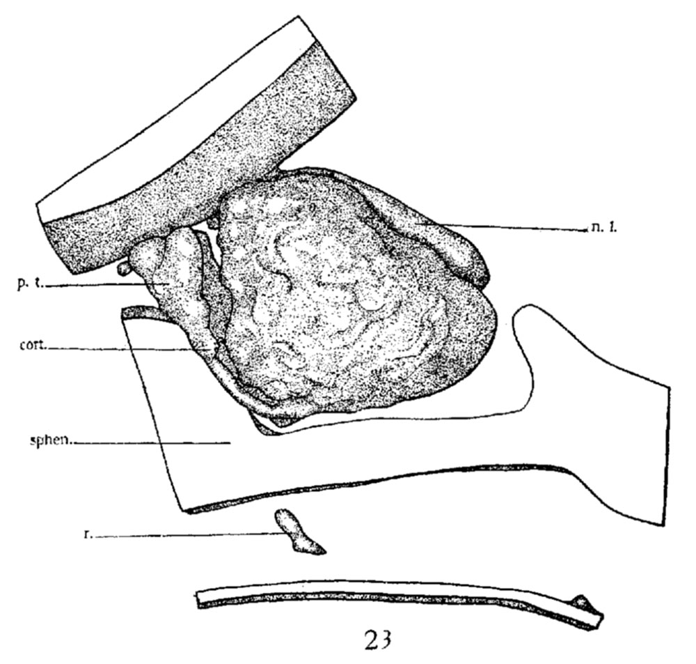
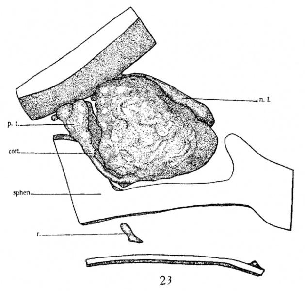
Fig. 23 Model of hypophysis region, eighteen-day embryo
viewed from left side. X 50. r., remains of stalk below sphenoid, or ‘pharyngeal hypophysis ’ Other abbreviations as in figure 22.
| Historic Disclaimer - information about historic embryology pages |
|---|
 |
See also Atwell WJ. The development of the hypophysis cerebri in man, with special reference to the pars tuberalis. (1926) Amer. J Anat. 37: 139-193.
Links: Pituitary Development | Rabbit Development
Reference
Atwell WJ. The development of the hypophysis cerebri of the rabbit (Lepus Cuniculus L.). (1918) Amer. J Anat. 24(2): 271-337
Cite this page: Hill, M.A. (2024, April 17) Embryology Atwell1918 fig23.jpg. Retrieved from https://embryology.med.unsw.edu.au/embryology/index.php/File:Atwell1918_fig23.jpg
- © Dr Mark Hill 2024, UNSW Embryology ISBN: 978 0 7334 2609 4 - UNSW CRICOS Provider Code No. 00098G
File history
Click on a date/time to view the file as it appeared at that time.
| Date/Time | Thumbnail | Dimensions | User | Comment | |
|---|---|---|---|---|---|
| current | 18:36, 14 November 2016 | | 1,000 × 953 (123 KB) | Z8600021 (talk | contribs) | |
| 18:36, 14 November 2016 | 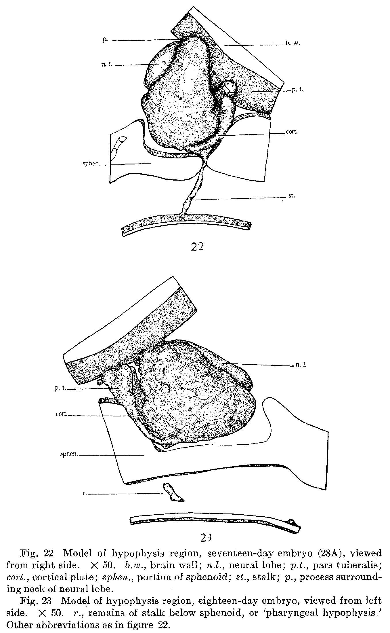 | 1,356 × 2,198 (465 KB) | Z8600021 (talk | contribs) |
You cannot overwrite this file.
File usage
The following page uses this file: