File:Arey1924 fig419.jpg
Arey1924_fig419.jpg (700 × 493 pixels, file size: 25 KB, MIME type: image/jpeg)
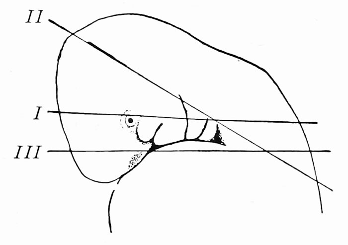
Fig. 419. Diagram to illustrate the planes of section made in dissecting the tongue
The development of the tongue may be studied from dissections of pig embryos 6, 9, and 13 mm. long. As the pharynx is bent nearly at right angles, it is necessary to cut away its roof by two pairs of sections passing in different planes. The first plane of section cuts through the eye and first two branchial arches just above the cervical sinus (Fig. 419, 1 ). From the surface, the razor blade should be directed obliquely dorsad in cutting toward the median line. Cuts in this plane should be made from either side. In the same way, make sections on each side in a plane forming an obtuse angle with the first section and passing dorsal to the cervical sinus {u). Now sever the remaining portion of the head from the body by a transverse section in a plane parallel to the first {uI). Place the ventral portion of the head in a watch glass of alcohol, and, under the dissecting microscope, remove that part of the preparation cranial to the mandibular arches. Looking down upon the floor of the pharynx, remove any portions of the lateral pharyngeal wall which may still interfere with a clear view of the pharyngeal arches, as seen in Fig. 84. Permanent mounts of the three stages mentioned above may be made and used for study by the student.
| Historic Disclaimer - information about historic embryology pages |
|---|
| Pages where the terms "Historic" (textbooks, papers, people, recommendations) appear on this site, and sections within pages where this disclaimer appears, indicate that the content and scientific understanding are specific to the time of publication. This means that while some scientific descriptions are still accurate, the terminology and interpretation of the developmental mechanisms reflect the understanding at the time of original publication and those of the preceding periods, these terms, interpretations and recommendations may not reflect our current scientific understanding. (More? Embryology History | Historic Embryology Papers) |
Reference
Arey LB. Developmental Anatomy. (1924) W.B. Saunders Company, Philadelphia.
Cite this page: Hill, M.A. (2024, April 24) Embryology Arey1924 fig419.jpg. Retrieved from https://embryology.med.unsw.edu.au/embryology/index.php/File:Arey1924_fig419.jpg
- © Dr Mark Hill 2024, UNSW Embryology ISBN: 978 0 7334 2609 4 - UNSW CRICOS Provider Code No. 00098G
File history
Click on a date/time to view the file as it appeared at that time.
| Date/Time | Thumbnail | Dimensions | User | Comment | |
|---|---|---|---|---|---|
| current | 12:35, 23 October 2016 |  | 700 × 493 (25 KB) | Z8600021 (talk | contribs) | |
| 12:34, 23 October 2016 | 1,602 × 571 (53 KB) | Z8600021 (talk | contribs) | ==Fig. 419. Diagram to illustrate the planes of section made in dissecting the tongue== {{Arey1924 Footer}} Category:Pig |
You cannot overwrite this file.
File usage
The following 2 pages use this file:


