File:Angle1918 plate17.jpg
From Embryology

Size of this preview: 364 × 600 pixels. Other resolution: 788 × 1,298 pixels.
Original file (788 × 1,298 pixels, file size: 348 KB, MIME type: image/jpeg)
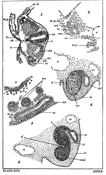
Plate XVII
Fig. 1. Cross section form the proximal end of the Wolffian body of an embryo 2.5 mm.long. III—4 xX 100.
Fig. 2. Left side of figure 1 more highly magnified I—5 x 190.
Fig. 3. An oblique section passing through the distal end of a 3 mm. embryo. The Wolffian duct and three Wolffian vesicles areshown. III—5 X 280.
Figs. 4 and 5. Cross sections from the distal end of a 4 mm. embryo. I—5 X 190.
Reference
Angle EJ. Development of the Wolffian Body in Sus Scrofa Domesticus.(1918) Trans. Amer. Micro. Soc. 37(4): 215-238.
File history
Click on a date/time to view the file as it appeared at that time.
| Date/Time | Thumbnail | Dimensions | User | Comment | |
|---|---|---|---|---|---|
| current | 15:15, 18 March 2019 |  | 788 × 1,298 (348 KB) | Z8600021 (talk | contribs) | Angle1918 |
You cannot overwrite this file.
File usage
The following page uses this file: