File:Ananomy of Ear.JPG
Ananomy_of_Ear.JPG (566 × 405 pixels, file size: 23 KB, MIME type: image/jpeg)
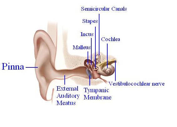
The image shows the anatomy of adult human ear:
Outer ear: Pinna, Eaxternal auditory meatus and tympanic membrane
Middle ear: Ossicles - Malleus, Incus and Stapes
Inner ear: Cochlea containing the Organ of Corti and Semi-circular canals containing semi-circular ducts
Image modified from <pubmed>20624897</pubmed>
This article is distributed under the terms of an Attribution–Noncommercial–Share Alike–No Mirror Sites license for the first six months after the publication date (see http://www.rupress.org/terms). After six months it is available under a Creative Commons License (Attribution–Noncommercial–Share Alike 3.0 Unported license, as described at http://creativecommons.org/licenses/by-nc-sa/3.0/).
I (z3333794) grant the public the non-exclusive right to copy, distribute, or display the Work under a Creative Commons Attribution-Noncommercial-Share Alike 3.0 Unported license, as described at http://creativecommons.org/licenses/by-nc-sa/3.0/ and http://creativecommons.org/licenses/by-nc-sa/3.0/legalcode."
File history
Click on a date/time to view the file as it appeared at that time.
| Date/Time | Thumbnail | Dimensions | User | Comment | |
|---|---|---|---|---|---|
| current | 12:11, 19 September 2012 |  | 566 × 405 (23 KB) | Z3333794 (talk | contribs) | The image shows the anatomy of adult human ear: Outer ear: Pinna, Eaxternal auditory meatus and tympanic membrane Middle ear: Ossicles - Malleus, Incus and Stapes Inner ear: Cochlea containing the Organ of Corti and Semi-circular canals containing semi |
You cannot overwrite this file.
File usage
The following file is a duplicate of this file (more details):
There are no pages that use this file.
