File:Amin1914 fig02.jpg
Original file (1,000 × 681 pixels, file size: 152 KB, MIME type: image/jpeg)
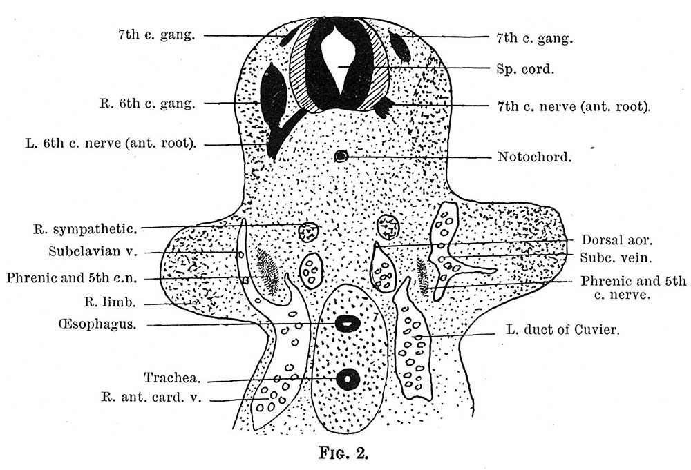
Fig. 2.
As the nerve is traced downwards, it passes ventrally, and comes to lie behind the anterior cardinal vein; and at a still lower level it establishes a communication with the fifth cervical nerve, and comes to lie in a cup-shaped invagination at the junction of the anterior cardinal vein with the primitive subclavian vein from the limb bud (fig. 2).
Phrenic nerve origin, course, and termination in a 6 mm embryo.
| Historic Disclaimer - information about historic embryology pages |
|---|
 |
Reference
Amin M. The course of the phrenic nerve in the embryo. (1914) J Anat Physiol. 48(2): 215-8. PMID 17232992
Cite this page: Hill, M.A. (2024, April 24) Embryology Amin1914 fig02.jpg. Retrieved from https://embryology.med.unsw.edu.au/embryology/index.php/File:Amin1914_fig02.jpg
- © Dr Mark Hill 2024, UNSW Embryology ISBN: 978 0 7334 2609 4 - UNSW CRICOS Provider Code No. 00098G
File history
Click on a date/time to view the file as it appeared at that time.
| Date/Time | Thumbnail | Dimensions | User | Comment | |
|---|---|---|---|---|---|
| current | 13:50, 23 January 2016 | | 1,000 × 681 (152 KB) | Z8600021 (talk | contribs) | {{Amin1914 figures}} |
You cannot overwrite this file.
File usage
The following 2 pages use this file: