File:Adrenal gland.png
Adrenal_gland.png (313 × 523 pixels, file size: 90 KB, MIME type: image/png)
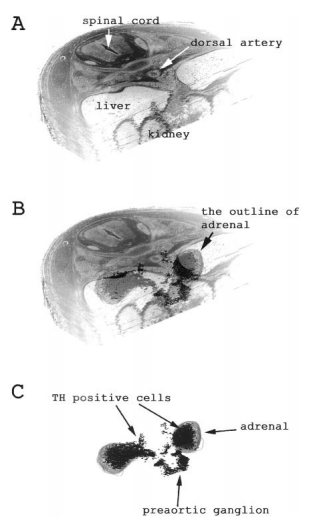
3D images showing TH - positive cells accumulating at the cranial and audal ends of the adrenal gland. A. A observation of adrenal gland and surrounding structures (at 17 days of gestation). B. Enhanced image to further focus on the dark TH - positive cells. C. Image with the other structures removed
Copyright: Open access Except as otherwise specifically noted, the articles with the Creative Commons icons in the journal are applied Creative Commons License (Creative Commons Attribution (CC BY) license, Creative Commons Attribution Non-Commercial (CC BY-NC) license and Creative Commons Attribution Non-Commercial-NoDerivs License (CC BY-NC-ND) etc.) by the publisher. (Due to constraints on the service or the system, some articles don't indicate the icons on the article page even if they are open access.)
- Note - This image was originally uploaded as part of an undergraduate science student project and may contain inaccuracies in either description or acknowledgements. Students have been advised in writing concerning the reuse of content and may accidentally have misunderstood the original terms of use. If image reuse on this non-commercial educational site infringes your existing copyright, please contact the site editor for immediate removal.
Mark Hill (talk) 12:54, 4 November 2018 (AEDT) Image relevant to project page. Not all required information included here on summary section, missing link to reference (added below). Should also include which species the study has been carried out in and included the reference in the figure legend on the project page, as well as in the associated text.
Yamamoto M, Yanai R & Arishima K. (2004). Study of migration of neural crest cells to adrenal medulla by three-dimensional reconstruction. J. Vet. Med. Sci. , 66, 635-41. PMID: 15240937
File history
Click on a date/time to view the file as it appeared at that time.
| Date/Time | Thumbnail | Dimensions | User | Comment | |
|---|---|---|---|---|---|
| current | 20:57, 15 October 2018 | | 313 × 523 (90 KB) | Z5112688 (talk | contribs) | 3D images showing TH - positive cells accumulating at the cranial and audal ends of the adrenal gland. A. A observation of adrenal gland and surrounding structures (at 17 days of gestation). B. Enhanced image to further focus on the dark TH - positive... |
You cannot overwrite this file.
File usage
The following file is a duplicate of this file (more details):
The following 2 pages use this file: