File:Adherens Junction 01.jpg
From Embryology

Size of this preview: 522 × 600 pixels. Other resolution: 696 × 800 pixels.
Original file (696 × 800 pixels, file size: 117 KB, MIME type: image/jpeg)
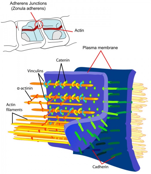
Adherens Junction
- actin microfilaments anchor the plaque that occurs under the membrane of each cell
- plaques not as dense also occur as hemi-form
- heart muscle, layers covering body organs, digestive tract
- transmembrane proteins
- Cadherin
- clustered as focal adhesion
Cadherin isoform naming was based upon the site of original identification: P-cadherin (placenta cadherin, cadherin 3), E-cadherin (epithelial cadherin cadherin 1), N-cadherin (neural cadherin, cadherin 2).
- Adhesion EM Images: GIT epithelia EM1 | GIT epithelia EM2 | GIT epithelia EM3 | Desmosome EM
- Adhesion Cartoons: Tight junction | Adherens Junction | Desmosome | Gap Junction
Cite this page: Hill, M.A. (2024, April 25) Embryology Adherens Junction 01.jpg. Retrieved from https://embryology.med.unsw.edu.au/embryology/index.php/File:Adherens_Junction_01.jpg
- © Dr Mark Hill 2024, UNSW Embryology ISBN: 978 0 7334 2609 4 - UNSW CRICOS Provider Code No. 00098G
File history
Click on a date/time to view the file as it appeared at that time.
| Date/Time | Thumbnail | Dimensions | User | Comment | |
|---|---|---|---|---|---|
| current | 12:25, 3 March 2012 |  | 696 × 800 (117 KB) | Z8600021 (talk | contribs) | ==Adherens Junction== * actin microfilaments anchor the plaque that occurs under the membrane of each cell * plaques not as dense also occur as hemi-form * heart muscle, layers covering body organs, digestive tract * transmembrane proteins * Cadherin * cl |
You cannot overwrite this file.