File:Adaptation Cerebellum.png
From Embryology

Size of this preview: 800 × 501 pixels. Other resolution: 2,918 × 1,828 pixels.
Original file (2,918 × 1,828 pixels, file size: 2.71 MB, MIME type: image/png)
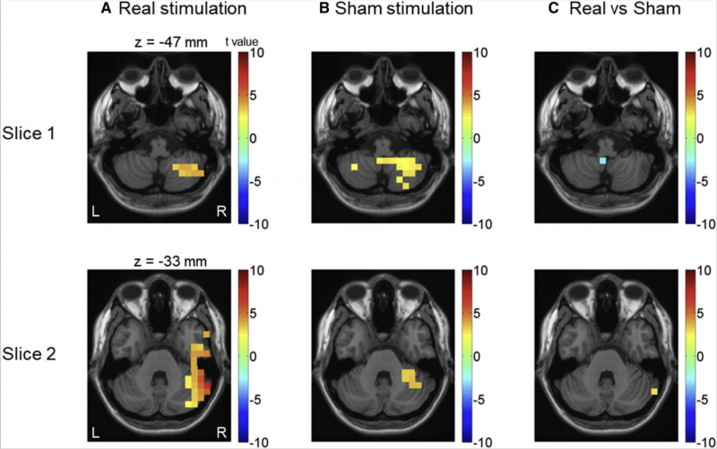
In real stimulation, a considerable increase in pre-stimulus low-frequency activity is only found in the right cerebellar hemisphere (near to the stimulation site). But in sham stimulation, pre-stimulus low-frequency activity increases in both the right cerebellar hemisphere and cerebellar vermis.[1].
Reference
- ↑ <pubmed>PMC5571438</pubmed>
Copyright
This is an open access article under the terms of the Creative Commons Attribution License, which permits use, distribution and reproduction in any medium, provided the original work is properly cited
- Note - This image was originally uploaded as part of an undergraduate science student project and may contain inaccuracies in either description or acknowledgements. Students have been advised in writing concerning the reuse of content and may accidentally have misunderstood the original terms of use. If image reuse on this non-commercial educational site infringes your existing copyright, please contact the site editor for immediate removal.
File history
Click on a date/time to view the file as it appeared at that time.
| Date/Time | Thumbnail | Dimensions | User | Comment | |
|---|---|---|---|---|---|
| current | 10:08, 26 October 2017 | | 2,918 × 1,828 (2.71 MB) | Z5076158 (talk | contribs) | In real stimulation, a considerable increase in pre-stimulus low-frequency activity is only found in the right cerebellar hemisphere (near to the stimulation site). But in sham stimulation, pre-stimulus low-frequency activity increases in both the righ... |
You cannot overwrite this file.
File usage
The following 2 pages use this file: