File:Abnormal formation of fertilisation cones .jpg
Abnormal_formation_of_fertilisation_cones_.jpg (256 × 256 pixels, file size: 5 KB, MIME type: image/jpeg)
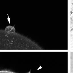
Heparin induces polyspermy and abnormal formation of fertilization cones
Formation of the fertilization cone was monitored in living eggs after sperm addition. (A) F-actin was visualized by microinjection of fluorescent phalloidin. (B) Transmission photomicrographs of the same eggs. Both phalloidin-stained actin networks and the transmission photomicrographs displayed formation of single and round fertilization cone (arrow) in the control egg by 9 min after adding sperm. In contrast, heparin-treated eggs displayed abnormal formation of multiple and piriform-shaped fertilization cones (arrowheads).
(Original figure legend, image based on data PMID 18974786)
Reference
<pubmed>18974786</pubmed>|[1]
Copyright
Copyright Puppo et al. This is an open-access article distributed under the terms of the Creative Commons Attribution License, which permits unrestricted use, distribution, and reproduction in any medium, provided the original author and source are credited.
- Note - This image was originally uploaded as part of an undergraduate science student project and may contain inaccuracies in either description or acknowledgements. Students have been advised in writing concerning the reuse of content and may accidentally have misunderstood the original terms of use. If image reuse on this non-commercial educational site infringes your existing copyright, please contact the site editor for immediate removal.
File history
Click on a date/time to view the file as it appeared at that time.
| Date/Time | Thumbnail | Dimensions | User | Comment | |
|---|---|---|---|---|---|
| current | 14:33, 14 August 2015 | | 256 × 256 (5 KB) | Z5015752 (talk | contribs) | Original file name: tileshop.fcgi.jpg PMID: 18974786 |
You cannot overwrite this file.
File usage
There are no pages that use this file.