File:1918 influenza virus virions EM.jpg

Original file (700 × 743 pixels, file size: 82 KB, MIME type: image/jpeg)
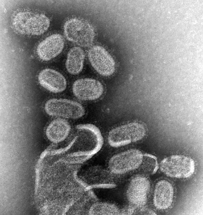
1918 Influenza Virus Virions EM
This negative stained transmission electron micrograph (TEM) shows recreated 1918 influenza virions that were collected from supernatants of 1918-infected Madin-Darby Canine Kidney (MDCK) cells cultures 18 hours after infection.
To separate these virions, the MDCK cells are spun down (centrifugation), and the 1918 virus in the fluid is immediately fixed for negative staining. The solid mass in lower center contains MDCK cell debris that did not spin down during the procedure. See PHIL 11098 for a colorized version of this image.
Dr. Terrence Tumpey, one of the organization’s staff microbiologists and a member of the National Center for Infectious Diseases (NCID), recreated the 1918 influenza virus in order to identify the characteristics that made this organism such a deadly pathogen. Research efforts such as this, enables researchers to develop new vaccines and treatments for future pandemic influenza viruses.
The 1918 Spanish flu epidemic was caused by an influenza A (H1N1) virus, killing more than 500,000 people in the United States, and up to 50 million worldwide. The possible source was a newly emerged virus from a swine or an avian host of a mutated H1N1 virus. Many people died within the first few days after infection, and others died of complications later. Nearly half of those who died were young, healthy adults. Influenza A (H1N1) viruses still circulate today after being introduced again into the human population in the 1970s.
Image name: 8160_lores.jpg http://phil.cdc.gov/PHIL_Images/8160/8160_lores.jpg
Content Providers(s): CDC/ Dr. Terrence Tumpey Photo Credit: Cynthia Goldsmith
Copyright Restrictions: None - This image is in the public domain and thus free of any copyright restrictions. As a matter of courtesy we request that the content provider be credited and notified in any public or private usage of this image.
File history
Click on a date/time to view the file as it appeared at that time.
| Date/Time | Thumbnail | Dimensions | User | Comment | |
|---|---|---|---|---|---|
| current | 11:29, 7 November 2011 | | 700 × 743 (82 KB) | S8600021 (talk | contribs) | ==1918 Influenza Virus Virions EM== This negative stained transmission electron micrograph (TEM) shows recreated 1918 influenza virions that were collected from supernatants of 1918-infected Madin-Darby Canine Kidney (MDCK) cells cultures 18 hours after |
You cannot overwrite this file.
File usage
The following page uses this file: