Category:EFIC
From Embryology
This Embryology category shows files and media listed below relate to the technique of Episcopic Fluorescence Image Capture (EFIC). A microscopic imaging technique that serially sections embedded biological specimens and photographs the tissue autofluorescence (epifluorescence) from the block surface. This generates an in register 2D image stack. Technique was described for the mouse in 2002 (PMID 11743576).
Pages in category 'EFIC'
The following 7 pages are in this category, out of 7 total.
Media in category 'EFIC'
The following 45 files are in this category, out of 45 total.
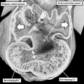
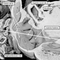
 Anderson2016-fig04.jpg 800 × 800; 99 KB
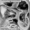
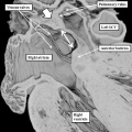
 Anderson2016-fig06.jpg 800 × 800; 123 KB
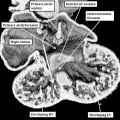
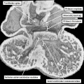
 Anderson2016-fig08a.jpg 800 × 800; 112 KB
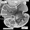
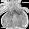
 Anderson2016-fig08b.jpg 800 × 800; 107 KB
 Anderson2016-fig09a.jpg 800 × 800; 106 KB
 Anderson2016-fig09b.jpg 800 × 800; 90 KB
 Anderson2016-fig12a.jpg 800 × 800; 120 KB
 Anderson2016-fig13a.jpg 800 × 800; 115 KB
 Anderson2016-fig17a.jpg 800 × 800; 98 KB
 Anderson2016-fig17b.jpg 800 × 800; 95 KB
 Anderson2016-fig24a.jpg 800 × 800; 107 KB
 Anderson2016-fig24b.jpg 800 × 800; 109 KB
 Anderson2016-fig25a.jpg 800 × 800; 122 KB
 Anderson2016-fig25b.jpg 800 × 800; 83 KB
 Anderson2016-fig26a.jpg 800 × 800; 126 KB
 Anderson2016-fig26b.jpg 800 × 800; 103 KB
 Anderson2016-fig27a.jpg 800 × 800; 117 KB
 Anderson2016-fig27b.jpg 800 × 800; 101 KB
 Anderson2016-fig28a.jpg 800 × 800; 117 KB
 Anderson2016-fig28b.jpg 800 × 800; 110 KB
 Anderson2016-fig29a.jpg 800 × 800; 111 KB
 Anderson2016-fig29b.jpg 800 × 800; 83 KB
 Anderson2016-fig31.jpg 800 × 800; 99 KB
 Anderson2016-fig32a.jpg 800 × 800; 108 KB
 Anderson2016-fig32b.jpg 800 × 800; 89 KB
 Anderson2016-fig33a.jpg 800 × 800; 110 KB
 Anderson2016-fig33b.jpg 800 × 800; 127 KB
 Anderson2016-fig34a.jpg 800 × 800; 105 KB
 Anderson2016-fig35a.jpg 800 × 800; 118 KB
 Anderson2016-fig38.jpg 800 × 800; 149 KB
 Anderson2016-fig39a.jpg 800 × 800; 86 KB
 Anderson2016-fig39b.jpg 800 × 800; 108 KB
 Anderson2016-fig40a.jpg 800 × 800; 102 KB
 Anderson2016-fig40b.jpg 800 × 800; 126 KB
 Anderson2016-fig42a.jpg 800 × 800; 90 KB
 Anderson2016-fig42b.jpg 800 × 800; 111 KB
 Anderson2016-fig46a.jpg 800 × 800; 100 KB
 Anderson2016-fig46b.jpg 800 × 800; 120 KB
 Heart outflow tract stage 14 01.jpg 2,039 × 996; 274 KB
 Heart outflow tract stage 14 02.jpg 996 × 996; 139 KB
 Heart outflow tract stage 14 03.jpg 989 × 996; 134 KB
 Stage16 EFIC C01.mp4 ; 1.09 MB
- Stage16 EFIC C02.mp4 ; 1.34 MB
- Stage16 EFIC S01.mp4 ; 1,018 KB
- Stage16 EFIC T01.mp4 ; 430 KB


