Category:Carnegie Stage 8
This Embryology category shows pages and media related to Carnegie stage 8 of embryonic development that occurs during Week 3 (post-fertilisation) or gestational age, GA, LMP week 5.
| Week: | 1 | 2 | 3 | 4 | 5 | 6 | 7 | 8 |
| Carnegie stage: | 1 2 3 4 | 5 6 | 7 8 9 | 10 11 12 13 | 14 15 | 16 17 | 18 19 | 20 21 22 23 |
| Carnegie Collection - Stage 8 | ||||||||||
|---|---|---|---|---|---|---|---|---|---|---|
| Serial No. | Grade | Fixative | Embedding Medium | Plane | Thinness (µm) | Stain | Year | Notes | ||
| 1399 | Poor | Formol | P | Transverse | 10 | (Stain - Haematoxylin Eosin) etc. | 1916 | "Mateer embryo" described by Streeter (1920a)[1] | ||
| 3412 | Poor | Formol | P | Transverse | 5-15 | Al. coch. E. au., or. | 1921 | |||
| 5960 | Good | Kaiserling | P | Transverse | 5 | Al. coch. & eosin | 1929 | Heuser (1932b)[2] | ||
| 6630 | Poor | Formol | P | Oblique | 6 | (Stain - Haematoxylin Eosin) | 1932 | |||
| 6815 | Poor | Formol | P | Oblique | 10 | Al. coch., or. G | 1933 | |||
| 7170a and b 7545 | Poor | Alc. | C-P | Transverse | 6 | (Stain - Haematoxylin Eosin) | 1935 | Twins | ||
| 7568 | Poor | Formoi | C-P | Transverse | 6 | (Stain - Haematoxylin Eosin) | 1938 | |||
| 7640 | Good | Formol & Bouin | P | Transverse | 10 | (Stain - Haematoxylin Eosin) | 1939 | George (1942)[3] | ||
| 7666 | Exc. | Formol-chrom. subl. | C-P | Transverse | 6 | (Stain - Haematoxylin Eosin) | 1939 | "H. 1515" | ||
| 7701 | Exc. | ? | C-P | Transverse | 8 | (Stain - Haematoxylin Eosin) | 1939 | |||
| 7822 | Good | Formoi | C-P | Transverse | 10 | (Stain - Haematoxylin Eosin) | 1940 | |||
| 7949 | Good | Zenker | p | Sagittal | 10 | (Stain - Haematoxylin Eosin) etc. | 1941 | |||
| 7972 | Good | Alc. & Bouin | C-P | Sagittal | 6 | (Stain - Haematoxylin Eosin) | 1942 | |||
| 8255 | Exc. | Bouin | C-P | Sagittal | 8 | (Stain - Haematoxylin Eosin), phlox. | 1944 | Slides showing embryo returned to Dr. Patten in 1962 | ||
| 8320 | Good | Formol | C-P | Sagittal | 8 | (Stain - Haematoxylin Eosin), phlox. | 1945 | |||
| 8352 | Good | Formol | C-P | Transverse | 8 | (Stain - Haematoxylin Eosin), phlox. | 1946 | |||
| 8371 | Poor | Alc. & Bouin | C-P | Sagittal | 8 | (Stain - Haematoxylin Eosin), phlox. | 1946 | |||
| 8671 | Exc. | Alc. & Bouin | C-P | Sagittal | 6 | (Stain - Haematoxylin Eosin), phlox. | 1949 | |||
| 8725 | Exc. | Alc. & Bouin | C-P | Sagittal | 6 | (Stain - Haematoxylin Eosin), phlox. | 1949 | Preparation method described by Heard (1957)[4] | ||
| 8727 | Exc. | Alc. & Bouin | C-P | Transverse | 8 | (Stain - Haematoxylin Eosin), phlox. | 1949 | Germ disc folded, possibly double (Hertig, 1968, fig. 180)[5] | ||
| 8820 | Good | Zenker-formol | ? | Transverse | 10 | (Stain - Haematoxylin Eosin) | 1951 | "Jones-Brewer I" (H. 1459) described by Jones and Brewer (1941)[6] | ||
| 9009a and b 9123 | Good | Formol | C-P | Sagittal | 6 | (Stain - Haematoxylin Eosin) | 1952 | Twins described briefly by Heuser (1954)[7] | ||
| 8371 | Good | Formol | C-P | Sagittal | 6 | (Stain - Haematoxylin Eosin) | 1953 | |||
| 9251 | Good | ? | C-P | Sagittal | 10-12 | Azan, H. & phlox. | 1954 | |||
| 9286 | Exc. | Formol | C-P | Transverse | 8 | Azan | 1955 | |||
| 10157 | Exc. | Formol | C-P | Transverse | ? | Cason | 1967 | |||
| 10174 | Exc. | Bouin | p | Transverse | 8 | Cason | 1967 | |||
Abbreviations
| ||||||||||
References
| ||||||||||
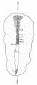
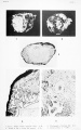
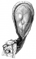
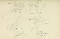
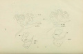
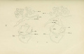
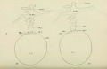
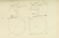
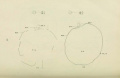
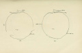
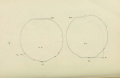
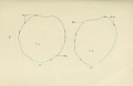
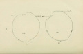
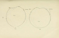
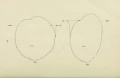
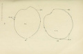
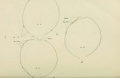
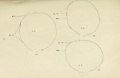
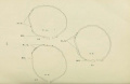
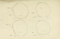
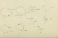
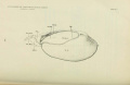
An historic example of this stage is shown by the Dobbin embryo, described in detail by the series of papers by Hill and Florian (1931a,b,c).[1][2][3] Abortion. Chorion, 11.5 x 8.5 x 4.5 mm. Chorionic cavity, 9 x 5.5 x 2.5 mm. Embryonic disc (narrow type), 0.96 x 0.41 mm. Primitive streak, 0.42 mm to notopore. Notochordal process, 0.42 mm. Notochordal canal communicates with cavity of umbilical vesicle by seven openings, the caudalmost of which is the ventral opening of a very short neurenteric canal. Prechordal plate, 0.03 mm; rostral end of notochordal process was at first mistaken for prechordal plate.
- ↑ Hill, J. P., and Florian, J. 1931a. The Development of Head-Process and Prochordal Plate in Man J Anat. 1931 Jan;65(Pt 2):242-6. PMID 17104317
- ↑ Hill, J. P., and Florian, J. 1931b. A Young Human Embryo (Embryo Dobbin) with Head-Process and Prochordal Plate. Phil. Tran. Roy. Soc. London B, 219, 443-486.
- ↑ Hill, J. P., and Florian, J. 1931c. Further note on the pro-chordal plate in man. J. Anat., 46, 46-47. PMID 17104356
Embryo Examples
Based on O'Rahilly, R. and Müller (1987)[1] and listed in order of length of notochordal process.
- Pha XVII. Chorionic cavity, 3.317 x 2.79 x 0.714mm. Embryonic disc, 0.412 mm. This embryo is said to resemble No. 7802 (stage 7). In addition to a notochordal process (Mazanec, 1959[2]., fig. 104) of 0.01 mm, however, it is thought to show probably the Anlage of the notochordal canal and an unusually large prechordal plate. Possible primordial germ cells were seen in the wall of the umbilical vesicle. Presumed age, 16-17 days. Median projection published (ibid., fig. 44).
- Carnegie No. 8820, Jones-Brewer I. Described by Jones and Brewer (1941)[3]. Hysterectomy. Chorionic cavity, 6 x 5 x 2.5 mm. Embryonic disc (broad type), 0.58 x 0.78 mm (in straight line); 0.6 x 0.79 mm (over curve). Primitive streak, 0.22 mm. Primitive node (0.06 mm) situated somewhat rostral to midpoint of embryonic disc. Three small, discontinuous cavities in node “represent the beginning of a neurenteric canal” which has no dorsal opening and does not communicate with the umbilical vesicle. Notochordal process, 0.0414 mm, but with no canalization. Hemocytoblasts and primitive erythroblasts identified in wall of umbilical vesicle (Bloom and Bartelmez, 1940). Presumed age, 18½ days. Dorsal and median projections published (Jones and Brewer, 1941, figs. 11 and 12; Mazanec, 1959[2], fig. 48).
- Carnegie No. 9286 Embryonic disc, 1.13 x 0.77 mm. Primitive streak, 0.38 mm. Notochordal process, 0.15 mm. An excellent specimen reconstructed by O'Rahilly and Müller (1981, fig. 3), who give details of ten other Carnegie specimens
- Carnegie No. 9009. Described briefly in an abstract by Heuser (1954). Hysterectomy. Monozygotic twin embryos. Embryonic discs, 0.9 and 0.66 mm. In each: primitive node in middle of disc, notochordal process with first evidence of canalization. Notochordal processes, 0.16 and 0.07 mm. Assigned to horizon VIII by Heuser, who estimated the age as 17 days. Reconstructed by O’Rahilly and Müller (1981, fig. 2B,C).
- Shaw. Described by Gladstone and Hamilton (1941)[4]. Hysterectomy. Chorion, 11 x 4.04 mm. Chorionic cavity, 8 x 3 mm. Embryonic disc (broad type), 1.05 x 1.34 mm. Notochordal process, 0.17 mm. Primitive pit and notochordal canal (which does not open into the umbilical vesicle). Prechordal plate identified but doubted by Mazanec (1959)[2]. No neural groove. Possible amniotic duct. Hemopoiesis (hemocytoblasts and primitive erythroblasts) under way in wall of umbilical vesicle and in connecting stalk. Chorionic villi and endometrium described by Hamilton and Gladstone (1942); trophoblast further described by Hamilton and Boyd (1960). Presumed age, 18 days. Median projection published (Gladstone and Hamilton, 1941; Mazanec, 1959[2], fig. 58).
- Wa 17 (Wagner). Described by Grosser (1931a,b).[5][6] Hysterectomy. Chorionic cavity, 8.5 x 8.5 x 7.5 mm. Embryonic disc (narrow type), 0.98 x 0.7 mm. Primitive streak, 0.5 mm. Notochordal process, 0.18 mm. Possible dorsal and ventral openings of the notochordal canal. Prechordal plate, 0.075 mm. Presumed age, about 19 days. Dorsal and median projections published (Grosser, 1931a, figs. 4 and 3; Hill and Florian, 1931b, fig. 7; Mazanec, 1959[2], fig. 59).
- Carnegie No. 8671. Low-power photomicrographs reproduced by Hertig (1968, figs. 47 and 181). Notochordal process, 0.23 mm.
- Kl. 13. Described by Grosser (1913). Traumatic abortion following salpingo-oophorectomy. Chorionic cavity, 8 x 6 mm. Embryonic disc, 0.67 x 0.5 mm. Primitive streak, 0.27 mm. Notochordal process, 0.2 mm. Notochordal canal, 0.25 mm (Florian, 1934c) with dorsal pit and ventral opening: notochordal plate intercalated in endoderm. Possible prechordal plate. Presumed age, about 18 days. Median projection published (Grosser, 1913, plate 27, fig. 4; Mazanec, 1959[2], fig. 62). Compared with other specimens by Grosser (1934).
- HEB-42. Described by Mazanec and Musilovà (1959). Curettage. Embryonic disc, 1.17 x 0.72 mm; 1.43 mm by flexible scale. Primitive streak, 0.54 mm. Primitive node, 0.06 mm. Primitive pit. Notochordal process, 0.25 mm. Small cavity (primordium of notochordal canal) in notochordal process. Prechordal plate not mentioned. Presumed age, 17-18 days. Dorsal and median projections published (ibid., figs. 1 and 2).
- Dy (Dyhrenfurth). Described by Triepel (1916)[7]. Abortion. Embryonic disc, 1.6 x 1.04 mm. Primitive streak, 0.11 mm. Notochordal process and plate, 0.3 mm. Primitive pit, neurenteric canal, and neural groove believed to be present. Anlagen of hypophysis and optic vesicles claimed unconvincingly (plane of section unsuitable). Embryonic disc bent ventrally through a right angle (normality of specimen questioned). First somite probably not present.
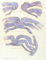
- 23 Somites Thompson and Brash (1923)[8] described a hysterectomy specimen that showed a chorionic cavity of 10 x 7.5 x 4 mm. Embryonic disc (broad type), 0.68 x 0.9 mm. Primitive streak and groove present. Notochordal process, 0.3 mm, with no distinct lumen but notochordal canal about to appear. Mazanec (1959) considered that, on the basis of the reconstruction, the notochordal process could not be more than 0.23 mm. Definite prechordal plate (Hill and Florian, 1931b). Rough dorsal and median drawings included (Thompson and Brash, 1923, figs, 2 and 3) and median projection published (Mazanec, 1959, fig. 55). This embryo belongs either to stage 7 or to stage 8.
- Schö (Schönholz). Described by Waldeyer (1929a,b)[9][10] Hysterectomy. Embryonic disc, 0.99 x 1.03 x 0.11 mm. Primitive streak, 0.51 mm, and node. Primitive groove and pit: “indentation” is perhaps “first Anlage of [notochordal] canal.” Notochordal process, 0.34 mm. Prechordal plate. Said to lie between Hugo (stage 7) and Peh. l-Hochstetter (stage 8). Dorsal and median projections published (ibid., figs. 6 and 5; Hill and Florian, 1931b, figs. 6 and 14; Mazanec, 1959[2], fig. 57).
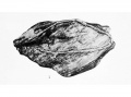
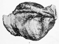
- Dobbin. Important specimen described in detail by Hill and Florian (1931a,b,c).[11][12][13] Abortion. Chorion, 11.5 x 8.5 x 4.5 mm. Chorionic cavity, 9 x 5.5 x 2.5 mm. Embryonic disc (narrow type), 0.96 x 0.41 mm. Primitive streak, 0.42 mm to notopore. Notochordal process, 0.42 mm. Notochordal canal communicates with cavity of umbilical vesicle by seven openings, the caudalmost of which is the ventral opening of a very short neurenteric canal. Prechordal plate, 0.03 mm; rostral end of notochordal process was at first mistaken for prechordal plate. Dorsal and median projections published (Hill and Florian, 1931b, figs. 1 and 2; Mazanec, 1959[2], fig. 60). The scale in fig. 3 of Hill and Florian (1931b) is incorrect (Florian, 1934c). Specimen is now housed in Hubrecht Laboratory, Utrecht (No. H91; HH 159).
- Carnegie No. 5960 Important specimen described by Heuser (1932b). Hysterectomy. Chorion, 15 x 14 x 9 mm. Embryonic disc (narrow type), 1.25 x 0.68 mm (in straight line); 1.53 x 0.75 mm (by flexible scale). Primitive streak, 0.5 (0.44?) mm. Primitive node, 0.2 (0.06?) mm, slightly caudal to midpoint of embryonic disc. Notochordal process, 0.42 mm (George, 1942). Notochordal canal (about 0.4 mm) opens ventrally. Prechordal plate, 0.15 mm. Florian (1934b), however, believed that the prechordal plate was situated further rostrally than shown by Heuser. Angiogenesis in umbilical vesicle, body stalk, and chorion. Angiogenesis in chorion described by Hertig (1935). Neural groove. Presumed age, 18 days. Dorsal and median projections published (Heuser, 1932b, figs. 33 and 47; Mazanec, 1959[2], fig. 63).
- Carnegie No. 7545. Embryonic disc, 1.52 x 1.03 mm. Primitive streak, 0.61 mm. Notochordal process, 0.43 mm. Reconstructed by O'Rahilly and Müller (1981, fig. 2E).
- Carnegie No. 7640. Described by George (1942). Tubal. Embryonic disc (broad type), 1.01 x 0.83 mm (in straight line); 1.16 mm by flexible scale. Primitive streak, node, and pit present. Notochordal process, 0.44 mm. Notochordal canal continuous with primitive pit; floor of canal has disappeared in its middle quarter. Prechordal plate, 0.12 mm, said to contain continuation of notochordal canal. Neural groove. Dorsal and median projections published (ibid., figs. 1 and 2).
- Peh. 1-Hochstetter (Peham). Described by Rossenbeck (1923). Chorion, 10 x 7.7 mm. Chorionic cavity, 6.8 x 5.3 mm. Embryonic disc (broad type; this might be disputed, however), 1.77 (Florian, 1934) x 1 mm. Primitive streak, 0.69 mm. Notochordal process (Mazanec, 1959[2], figs. 107 and 108), 0.6 mm. Notochordal canal ready to break through into umbilical vesicle in one section. Prechordal plate (confirmed by Florian, 1931), 0.08 mm. Indication of hindgut (Florian, 1934b). Allanto-enteric diverticulum (Florian, 1930a). Dorsal and median projections published (Hill and Florian, 1931b, figs. 9 and 16; Mazanec, 1959, fig. 61).
- R. S. (Robb Smith). Described by Odgers (1941)[14]. Hysterectomy. Embryonic disc (broad type), 1.5 x 1.36 mm. Primitive streak, 0.4 mm. Notochordal plate (intercalated in endoderm), 0.7 mm. Neurenteric canal extends vertically from amniotic cavity to umbilical vesicle. Prechordal plate, 0.29 mm, containing perhaps remains of notochordal canal. Commencing neural groove. Dorsal and median projections published (ibid., figs. 1 and 2).
- Western Reserve No. 1. Described by Ingalls (1918)[15]. Abortion. Chorion, 9.1 x 8.2 x 6.5 mm. Chorionic cavity, 8 x 7 x 5 mm. Embryonic disc (narrow type), 2 (1.87?) x 0.75 mm. Primitive streak, 0.67 mm. Primitive pit present. Notochordal process, 0.75 (0.65?), Hill and Florian, 1931b) mm. Notochordal canal, 0.34 mm, with three ventral openings into umbilical vesicle. Prechordal plate identified (but not 0.4 mm in length, according to Mazanec, 1959). Dorsal and median projections published (Hill and Florian, 1931b[12], figs. 10 and 17; Mazanec, 1959[2], fig. 64).
Precise measurements of the notochordal process have not been provided in the accounts of the following embryos.
- M’lntyre. Described by Bryce (1924)[16] and M’Intyre (1926). Hysterectomy. Chorion, 14 x 13 x 8 mm. Embryonic disc, 1.37 x 0.5 mm. Primitive streak, 0.32 mm. Notochordal plate, neurenteric canal, and prechordal plate (with cavity) present. Neural groove, future foregut, U-shaped pericardial cavity, and “some general resemblance to somites” noted. Rough dorsal and median drawings included (Bryce, 1924[16], figs. 5 and 49). May belong to stage 9.
- Frassi’s specimen (Keibel, 1907; Frassi, 1908) was illustrated as Normentafel No. 1 by Keibel and Elze (1908). Embryonic disc, 1.17 x 0.6 mm. Primitive streak and neurenteric canal identified. Neural groove. No somites.
- Gle., or Gläveke (von Spee, 1889 and 1896). Illustrated as Normentafel No. 2 by Keibel and Elze (1908). See also Kollmann (1907) and Keibel and Mall (1910, 1912). Chorion, 6 x 4.5 mm. Chorionic cavity, 5.3 x 3.8 mm. Embryonic disc, 1.54 mm. Primitive streak and node, notochordal plate, and neurenteric canal (Van Beneden, 1899) identified (Keibel and Mall, 1912, fig. 231). Neural groove. Indication of foregut and pericardial cavities. Anlage of endocardium. No somites detected, although a small cavity on the left side (Keibel and Elze, 1908, fig. 4f) could be considered as the first Anlage of a myocoele. May belong to stage 9.
- Strahl (1916)[17] described briefly a specimen that possessed a notochordal process and canal, and apparently a prechordal plate. A median drawing was included (ibid., fig. a) but measurements were not provided.
- Vuill., or Vulliet. Illustrated schematically by Eternod (1899a and 1909), Kollmann (1907), and Keibel and Mall (1912). Chorion, 10 x 8.2 x 6 mm. Chorionic cavity, 9 x 7.2 x 5 mm. Embryonic disc, 1.3 mm. Notochordal and neurenteric canals (Eternod, 1899b).
- Cordier and Coujard (1939) described an embryo of 1.05 mm showing a neural groove and folds, notochordal canal, primordial germ cells, but no somites, no intra-embryonic coelom, and no cardiac rudiment.
- Carnegie No. 8727. Photomicrograph reproduced by Hertig (1968, fig. 180). The partial duplication of the embryonic disc shown in this specimen would presumably have resulted in conjoined twins.
Certain other embryos that probably belong to stage 8.
- Krukenberg (1922) and Fitzgerald- Brewer I (Brewer and Fitzgerald, 1937).
- Boerner-Patzelt and Schwarzacher (1923) described an unsatisfactory specimen (embryonic disc, 0.47 x 0.43 mm) that showed a neurenteric canal,
- Broman (1936) described in detail an abnormal specimen (“Lqt”) in which the primitive streak showed “overgrowth” in relation to the neurenteric canal, resulting in a dislocation within the embryonic disc.
References
- ↑ O'Rahilly, R. and Müller, F. Developmental Stages in Human Embryos. Carnegie Institution of Washington Publication 637 (1987).
- ↑ 2.00 2.01 2.02 2.03 2.04 2.05 2.06 2.07 2.08 2.09 2.10 Mazanec, K. 1959. Blastogenese des Metjschen. Fischer, Jena.
- ↑ Jones, H. O., and Brewer, J. I. 1941. A human embryo in the primitive-streak stage (Jones-Brewer ovum I). Carnegie Instn. Wash. Publ. 525, Contrib. Embryol., 29, 157-165.
- ↑ W J Hamilton, R J Gladstone A presomite human embryo (Shaw) - the implantation. J. Anat.: 1942, 76(Pt 2);187-203 PMID 17104888
- ↑ Grosser, O. 1931a. Primitivstreifen und Kopffortsatz beim Menschen. Verb. Anat. Ges., Erg. Heft Anat. Anz., 71,135—139.
- ↑ Grosser, O. 1931c. Weiteres uber den Primitivstreifen des Menschen. Verb. Anat. Ges., Erg. Heft Anat. Anz., 72, 42-44.
- ↑ Triepel, H. 1916. Ein menschlicher Embryo mit Canalis neurentericus. Chordulation. Anat. Hefte, 54, 149-185.
- ↑ Thompson, P., Description of a Human Embryo of Twenty-three Paired Somites. J Anat Physiol: 1907, 41(Pt 3);159-71 PMID 17232726
- ↑ Waldeyer, A. 1929a. Ein junges menschliches Ei in situ (Schönholz), Z Anal Entw., 90, 412-457
- ↑ Waldeyer, A. 1929b. Mesodermbildung bei einem jungen menschlichen Embryo. Anat Anz., 35, 145-151.
- ↑ Hill, J. P., and Florian, J. 1931a. The Development of Head-Process and Prochordal Plate in Man J Anat. 1931 Jan;65(Pt 2):242-6. PMID 17104317
- ↑ 12.0 12.1 Hill, J. P., and Florian, J. 1931b. A Young Human Embryo (Embryo Dobbin) with Head-Process and Prochordal Plate. Phil. Tran. Roy. Soc. London B, 219, 443-486.
- ↑ Hill, J. P., and Florian, J. 1931c. Further note on the pro-chordal plate in man. J. Anat., 46, 46-47. PMID 17104356
- ↑ Odgers, P. N. B. 1941. A presomite human embryo with a neurenteric canal (embryo R. S.). J. Anat, 75, 381-388.
- ↑ Ingalls NW. A human embryo before the appearance of the myotomes. (1918) Contrib. Embryol., Carnegie Inst. Wash. No.23 Publ. 227, 7:111-134.
- ↑ 16.0 16.1 Bryce, T. H. 1924. Observations on the early development of the human embryo. Trans. Roy. Soc. Edinburgh, 53, 533— 567.
- ↑ Strahl, H. 1916. Uber einen jungen menschiichen Embryo nebst Bemerkungen zu C. Rabl's Gastrulationstheorie. Anat. Hefte, 54, 113-147.
Subcategories
This category has the following 30 subcategories, out of 30 total.
C
- Carnegie Embryo 10157
- Carnegie Embryo 10174
- Carnegie Embryo 1399
- Carnegie Embryo 3412
- Carnegie Embryo 5960
- Carnegie Embryo 6630
- Carnegie Embryo 6815
- Carnegie Embryo 7170a
- Carnegie Embryo 7545
- Carnegie Embryo 7568
- Carnegie Embryo 7640
- Carnegie Embryo 7666
- Carnegie Embryo 7701
- Carnegie Embryo 7822
- Carnegie Embryo 7949
- Carnegie Embryo 7972
- Carnegie Embryo 8255
- Carnegie Embryo 8320
- Carnegie Embryo 8352
- Carnegie Embryo 8371
- Carnegie Embryo 8671
- Carnegie Embryo 8725
- Carnegie Embryo 8727
- Carnegie Embryo 8820
- Carnegie Embryo 9009
- Carnegie Embryo 9251
- Carnegie Embryo 9286
Pages in category 'Carnegie Stage 8'
The following 58 pages are in this category, out of 58 total.
C
- Template:Carnegie Collection stage 8 table
- Template:Carnegie Embryo Stage8
- Carnegie stage 8
- Carnegie Stage 8 - "Dobbin" Embryo
- Template:Carnegie stage 8 links
- Template:CE10157
- Template:CE10174
- Template:CE1399
- Template:CE3412
- Template:CE5960
- Template:CE6630
- Template:CE6740
- Template:CE6815
- Template:CE7568
- Template:CE7640
- Template:CE7666
- Template:CE7701
- Template:CE7822
- Template:CE7949
- Template:CE7972
- Template:CE8255
- Template:CE8320
- Template:CE8352
- Template:CE8371
- Template:CE8671
- Template:CE8725
- Template:CE8727
- Template:CE8820
- Template:CE9009
- Template:CE9123
- Template:CE9251
- Template:CE9286
- Template:CS8
P
- Paper - A human embryo before the appearance of the myotomes (1918)
- Paper - A presomite human embryo (Shaw) - the implantation
- Paper - A presomite human embryo (Shaw) - the implantation (1942)
- Paper - A presomite human embryo (Shaw) with primitive streak and chorda canal with special reference to the development of the vascular system (1941)
- Paper - A presomite human embryo with a neurenteric canal (embryo R.S.)
- Paper - A Young Human Embryo (Embryo Dobbin) with Head-Process and Prochordal Plate
- Paper - Further note on the pro-chordal plate in man
- Paper - Normal development of early human embryos: Observation of 90 specimens at Carnegie stages 7 to 13
- Paper - The Development of Head-Process and Prochordal Plate in Man
R
Media in category 'Carnegie Stage 8'
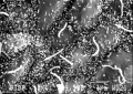
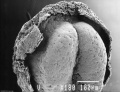
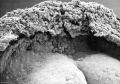
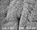
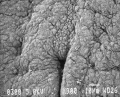
The following 81 files are in this category, out of 81 total.
 Gray0031.jpg 1,371 × 1,309; 270 KB
 HillFlorian1931a fig01.jpg 594 × 1,372; 74 KB
 HillH159 Stage 8 bf01.jpg 1,807 × 1,768; 237 KB
 HillH159 Stage 8 bf02.jpg 1,807 × 1,768; 264 KB
 HillH159 Stage 8 bf03.jpg 1,706 × 1,643; 168 KB
 HillH159 Stage 8 bf04.jpg 1,706 × 1,643; 214 KB
 Ingalls1918 plate 1 fig03.jpg 800 × 576; 72 KB
 Ingalls1918 plate 1 fig1+2.jpg 1,000 × 428; 81 KB
 Ingalls1918 plate 1 fig4+5.jpg 1,000 × 672; 161 KB
 Ingalls1918 plate 1.jpg 749 × 1,200; 164 KB
 Ingalls1918 plate 2 fig1+2.jpg 1,000 × 356; 50 KB
 Ingalls1918 plate 2 fig3+4.jpg 1,000 × 356; 50 KB
 Ingalls1918 plate 2 fig5+6.jpg 1,000 × 356; 60 KB
 Ingalls1918 plate 3 fig1.jpg 604 × 399; 17 KB
 Ingalls1918 plate 3 fig2.jpg 604 × 399; 21 KB
 Ingalls1918 plate 3 fig3.jpg 604 × 399; 25 KB
 Ingalls1918 plate 3 fig4.jpg 604 × 399; 22 KB
 Ingalls1918 plate 3 fig5.jpg 604 × 399; 19 KB
 Ingalls1918 plate 3.jpg 905 × 1,200; 132 KB
 Ingalls1918 plate 4 fig1.jpg 1,000 × 739; 92 KB
 Ingalls1918 plate 4 fig2.jpg 1,000 × 739; 151 KB
 Keiller Glaevecke embryo drawing 1.jpg 1,280 × 1,695; 484 KB
 Keiller Glaevecke embryo drawing 2.jpg 1,280 × 1,602; 403 KB
 Stage7 SEM4.jpg 450 × 332; 60 KB
 Stage8 bf1.jpg 659 × 913; 18 KB
 Stage8 bf10.jpg 1,000 × 515; 86 KB
 Stage8 bf11.jpg 1,000 × 515; 94 KB
 Stage8 bf2.jpg 544 × 800; 18 KB
 Stage8 bf3.jpg 640 × 1,000; 24 KB
 Stage8 bf4.jpg 600 × 449; 17 KB
 Stage8 bf5.jpg 500 × 656; 33 KB
 Stage8 bf6.jpg 500 × 656; 31 KB
 Stage8 bf7.jpg 668 × 1,000; 147 KB
 Stage8 bf8.jpg 657 × 1,000; 155 KB
 Stage8 bf9.jpg 1,000 × 515; 82 KB
 Stage8 cartoon 01.jpg 578 × 936; 145 KB
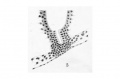
 Stage8 nodal cilia.jpg 450 × 321; 50 KB
 Stage8 SEM1.jpg 257 × 450; 25 KB
 Stage8 sem1.jpg 828 × 1,000; 109 KB
 Stage8 sem2.jpg 822 × 1,000; 142 KB
 Stage8 sem3.jpg 1,000 × 709; 91 KB
 Stage8 sem5.jpg 1,302 × 1,000; 113 KB
 Stage8 sem6.jpg 1,000 × 698; 87 KB
 Stage8 sem7.jpg 1,000 × 805; 139 KB
 Stage8 sem7a.jpg 800 × 644; 101 KB
 Stage8 sem7b.jpg 600 × 483; 65 KB
 Stage8 sem7c.jpg 400 × 322; 32 KB
 Turner1920 fig01-4.jpg 1,000 × 651; 53 KB
 Turner1920 fig05-6.jpg 1,000 × 651; 35 KB
 Turner1920 fig07-8.jpg 1,000 × 651; 43 KB
 Turner1920 fig09-10.jpg 1,000 × 651; 37 KB
 Turner1920 fig11-12.jpg 1,000 × 651; 47 KB
 Turner1920 fig13-14.jpg 1,000 × 651; 41 KB
 Turner1920 fig15-16.jpg 1,000 × 651; 47 KB
 Turner1920 fig17-18.jpg 1,000 × 651; 37 KB
 Turner1920 fig19-20.jpg 1,000 × 651; 41 KB
 Turner1920 fig21-22.jpg 1,000 × 651; 36 KB
 Turner1920 fig23-24.jpg 1,000 × 651; 38 KB
 Turner1920 fig25-26.jpg 1,000 × 651; 35 KB
 Turner1920 fig27-28.jpg 1,000 × 651; 39 KB
 Turner1920 fig29-30.jpg 1,000 × 651; 33 KB
 Turner1920 fig31-32.jpg 1,000 × 651; 33 KB
 Turner1920 fig33-34.jpg 1,000 × 651; 26 KB
 Turner1920 fig35-36.jpg 1,000 × 651; 29 KB
 Turner1920 fig37-38.jpg 1,000 × 651; 26 KB
 Turner1920 fig39-40.jpg 1,000 × 651; 27 KB
 Turner1920 fig41-42.jpg 1,000 × 651; 25 KB
 Turner1920 fig43-44.jpg 1,000 × 651; 28 KB
 Turner1920 fig45-46.jpg 1,000 × 651; 26 KB
 Turner1920 fig47-48.jpg 1,000 × 651; 30 KB
 Turner1920 fig49-50.jpg 1,000 × 651; 26 KB
 Turner1920 fig51-53.jpg 1,000 × 651; 32 KB
 Turner1920 fig54-56.jpg 1,000 × 651; 30 KB
 Turner1920 fig57-59.jpg 1,000 × 651; 31 KB
 Turner1920 fig60-62.jpg 1,000 × 651; 28 KB
 Turner1920 fig63-65.jpg 1,000 × 651; 30 KB
 Turner1920 fig66-68.jpg 1,000 × 651; 30 KB
 Turner1920 fig69-72.jpg 1,000 × 651; 36 KB
 Turner1920 fig73-81.jpg 1,000 × 651; 37 KB
 Turner1920 plate01.jpg 1,000 × 651; 33 KB
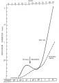 Yolk sac and amniotic cavity volume graph.jpg 719 × 1,000; 50 KB


