Book - Contributions to Embryology Carnegie Institution No.1-7
| Embryology - 18 Apr 2024 |
|---|
| Google Translate - select your language from the list shown below (this will open a new external page) |
|
العربية | català | 中文 | 中國傳統的 | français | Deutsche | עִברִית | हिंदी | bahasa Indonesia | italiano | 日本語 | 한국어 | မြန်မာ | Pilipino | Polskie | português | ਪੰਜਾਬੀ ਦੇ | Română | русский | Español | Swahili | Svensk | ไทย | Türkçe | اردو | ייִדיש | Tiếng Việt These external translations are automated and may not be accurate. (More? About Translations) |
Mall FP. On the fate of the human embryo in tubal pregnancy. (1915) Contrib. Embryol., Carnegie Inst. Wash. Publ. 221, 1: 1-104.
| Historic Disclaimer - information about historic embryology pages |
|---|
| Pages where the terms "Historic" (textbooks, papers, people, recommendations) appear on this site, and sections within pages where this disclaimer appears, indicate that the content and scientific understanding are specific to the time of publication. This means that while some scientific descriptions are still accurate, the terminology and interpretation of the developmental mechanisms reflect the understanding at the time of original publication and those of the preceding periods, these terms, interpretations and recommendations may not reflect our current scientific understanding. (More? Embryology History | Historic Embryology Papers) |
Bibliography and Plates
Bibliography of Papers Cited
ASCHOFF, Anatomie der Schwangcrschaft. Ziegler's Beitriige, Bd. 25, Jena, 1899.
BRYCE and TEACHER, Contributions to the study of the early development and imbedding of the human ovum. Glasgow, 1908.
FRASSI, Ueber ein junges menschliches Ei in situ. Archiv fur mikroskop. Anat. u. Entwicklungsgeschichte, Bd. 70, 1907.
GIACOMINI, Probleme aus Entwickelungsanomalien d. menschl. Embryo. Merkel and Bonnet, Anatomische Hefte, Bd. 4, 1904.
GROSSER, The development of the egg membranes and the placenta; menstruation. Keibel and Mall, Human Embryology, Vol. I, Chap. VII. Phila., 1910.
GROSSER, Eihaute und der Plazenta. Wien and Leipzig, 1909.
HERZOG, A contribution to our knowledge of the earliest known stages of placentation and embryonic development in man. Amer. Jour. Anat., Vol. IX. 1909.
HOFBAUER, Grundziige einer Biologic der menschlichen Plazenta. Wien und Leipzig, 1905.
HOFBAUER, Die menschliche Plazenta als Assimilationsorgan. Sammlung klinischer Vortrage, Leipzig. 1907.
JUNG, Ei-Einbettung beim menschlicheu Weibe. Berlin, 1908.
KEIBEL, Die aussere Korperform und der Entwickelungsgrad der Organe bei AfT enembryonen. Menschenaffen, Part IX, Wiesbaden, 1906.
KROEMER, Bau der menschlichen Tube. Leipzig, 1906.
MALL, A contribution to the study of the pathology of early human embryos. Welch Festschrift, Johns Hopkins Hospital Reports, vol. 9, 1900.
MALL, Second contribution to the study of the pathology of early human embryos. Vaughan Festschrift, Contributions to Medical Research, Ann Arbor, 1903.
MALL, A study of the causes underlying the origin of human monsters. (Third contribution to the study of the pathology of human embryos.) Jour, of Morphology, vol. 19, 1908. Also published as a monograph by the Wistar Institute of Anatomy, Phila., 1908.
MALL, The pathology of the human ovum. Keibel and Mall, Human Embryology, Vol. I, Chap. IX, Phila., 1910.
OPITZ, Ueber die Ursachen der Ansiedlung des Eies im Eileiter, Zeitschr. fiir Geburtshiilfe und Gynak., Bd. 48. Stuttgart, 1903.
PETERS, Die Einbettung des menschlichen Eies. Leipzig and Wien, 1899.
RETZIUS, Das Magma reticu!6 des menschlichen Eies. Biolog. Untersuehungen, Bd. I. Stockholm, 1890.
STRAHL and BENEKE, Ein junger menschlicher Embryo. Wiesbaden, 1910.
VALPEAC, Embryologie ou Ovologie humaine, contenant 1'histoire desscriptive et iconographique de I'truf human. Paris, 1833.
VEIT, Verschleppung der Chorionzotten. Wiesbaden, 1905.
VON WINCKEL, Ueber die menschl. Missbildungen, Samml. klin. Vortrage. Leipzig, 1904.
WALLGREN, Zur mikroskop. Anatomie der Tuberschwangerschaft beim Menschen, Anatom. Heft, Bd. 27. Wiesbaden, 1905.
WILLIAMS, Obstetrics, third edition. New York, 1912.
WERTH, Die Extrauterineschwangeschaft. Von Winckel's Handbuch der Geburtshulfe. Bd. 2, Teil. 2. Weissbaden, 1904.
Explanation of Plates
Plate 1
Fig. 1. Section through the attachment of the villi to the tube wall in No. 109, showing the extension of the trophoblast into the veins, with the destruction of their endothelial lining. X 72.
Fig. 2. Section through the veins in the tube wall of No. 109, showing a more extensive invasion of trophoblast than in the veins pictured in fig. 1. X 100.
Fig. 3. Section through an extension of vacuolated syncytium between the tips of the villi and the tube wall (No. 109). At B. V. a blood vessel is tapped and between it and the villus there is an extensive hemorrhage of blood into the spaces of the vacuolated syncytium. X 50.
Fig. 4. Syncytium covering a typical villus (No. 808). Stained with hematoxylin and aurantia. The blood corpuscles fill the spaces of the syncytium and fragments of corpuscles lie within the protoplasm of the cells. There are all gradations between complete blood corpuscles and granular protoplasm which take on the same color. X 300.
Plate 2
Fig. 1. A villus which is undergoing fibrous degeneration (No. 694). X 160.
Fig. 2. Section through the last remnant of the ovum in apposition with the tube wall (No. 472). The villi have undergone extensive degeneration and the trophoblast and part of the tube wall are hyaline. X 120.
Fig. 3.Tip of a fibrous villus from No. 670, partly covered with trophobhist, which extends over in the adjacent clot, where it is undergoing hyaline degeneration. All stages of trophoblast undergoing hyaline degeneration are shown. X 100.
Fig. 4.Section through a necrotic villus of No. 575 which is covered with an irregular mass of dead epithelium. At points the nuclear mass is clumped and somewhere the necrotic mass radiates between the blood cells. There are also accumulations of leucocytes. X 105.
Fig. 5. Section through an island of vacuolated syncytium which is extensively infiltrated with leucocytes (No. 567). X 55.
Fig. 6. Specimen No. 430, showing extreme degeneration of the syncytium. The protoplasm is converted into a hyaline mass and chromatin is represented as nuclear substance.
Fig. 7. Portion of diagram shown on page 80 (No. 670), enlarged 700 diameters. The invagination of the epithelial cells is very pronounced. There are also numerous large, protoplasm cells seen in the spaces of the mesenchyme. These are the so-called Hofbauer cells.
Plate 3
Fig. 1. Section through the point of attachment between a fibrous villus and the tube wall of No. 570. The epithelial covering has been detached and the space is filled with blood. X100.
Fig. 2. Section through the degenerated ovum in situ (No. 754). The coelom is filled with mottled granular magma into which there are radiating cells of mesenchyme. X 85.
Plate 4
FIG. 1. Section through a part of a l:u;^c viihiv fro n \'" inueoid degeneration of tin mesenchyme. /:', fpithelial CO ,'/, inesenchx me ; ,S', la
FIG. 2. Section through the ovum (No an extensive magma within the coelom. > 75.
Kir,. 3. Section through the periphery of a blood clol wandering trophobl;
a small villus in Nu. iny.
Plate 5
FIG. 2. Section through the blood clot and chorion of No. 396 which contains two bodir ly the remnant of the embryo, E, and the other, (.'. r.,"i the umbilical vesicle. > 35.
Photograph of section through the attachment of the embry . Merit to tin- clu.nonic \vall of
No. 342. The rudiment which is represented by a. ck-nse mass of cells lyini; within a cavity upon a pedicle which is probably the degenerated umbilical cord. At its attachment to the choi ion there are indications of blood vfsst-K.
Section through the remnant of the ovum within the tube of Xo. 29S. Only a few tihmus villi i<
Section through the degenerated chorion of No. 4.SS. The wall has been Si injured in the
manipulation re.|uirerl to remove u from tin- tube lumen. The firlnm is filled with an magma in which there are numerous membrane-like bodies which may re] ryo. X 36.
Fie. 1 drawn hy IX^'Hiv Ivters; I-"ii.' 4 drnwn by J K. Uidusch
Plate 6
Fig. 1. Retouched photograph of embryo. No. 25ft. The small openings over the eyes represent the holes from which the brain has escaped. X 5%.
Fig. 2. Photograph of of No. 612 o kink on one side and th arhich contains the Fallopian tube. Direction of the section is also shown. X 1.
Fig. 3. Photograph of specimen. Xo. 495. si al 01 the tube ami direction .1 .1 ., -lie block.
Fig. 4. Photograph ., X 2.
Fig. 5. Photograph of specimen, Xo. 602. shou-ir 'ion of the X 1.
Fig. 6. I 1 h of the blocks cut from the d . alpin^itis ma\ ot. XI.
Fig. 7.i if the section. There is a very b X 1.
Plate 7
Fn. 1. The tn be nf N<>. 657 which has been dissected so as to show the embryo in position. The sp.-irf 1 nion
and the tube wall is filled with a partly organized clot. The embryo appears to be normal. X r > ,-,. FIG. i. Section through a degenerated villus of No. 575 lying within the blood clot. It may be noted that the fibrin of
the blood clot does not touch the villus. Numerous leucocytes are also shown. X45. Km. 3. Photograph of specimen No. 575. FIG. 4. Retouched photograph of a villus undergoinj; mucoid degeneration, \o. 657. It is partly infiltrated with
1< m-m.-ytes. X about 65. Fn,. 5. Photograph of the collapsed ovum within organized clot surrounded with great masses of leucocytes i No.
The crjelom is rilled with dense magma. X about 40.
Plate 8
Fig. 1. Outline of tube showing distention caused by the pregnancy (No. 720) X l
Fig. 2. Pus tube opposite the pregnancy iti No. 7+1.
Fig. 3. Photograph of degem > idiati . M.nti a tip of villus in No. 741. ( )n ritiirr side there are la . s. X 55.
Fig. 4. Semidiagrammati urn within the tube of No. 741. Tlic ovum and villi are drawn in black. The red blood clots are striated and I o white. X 2.
Fig. 5. Photograph of transverse . with extensive t'ollicula' I No. 726). X about 15.
Fig. 1 (734)
Fig. 2 (734)
F,g. 4 (825)
FIG. 1. Semidiagrammatic sketch of the collapsed ovum within the tube (No. < \-\:m and the
villi are black. The red blood is i is white.
FIG. 2. Photograph of transverse section of the tube (No. 734), showing i lumen. X H.
l ; ir,. 3. Outline sketch of a transve
The ovum and villi an
l>v broken lines, and thi < 4.
FIG. 4. Tube sliou-ini; extensi '
is an indii-ai ion of i upture. Xai
1- IG. .X Seetion throu] i'
ovum, tin- ca of whi i no
Fig. 1 (775)
Fig. 3 (729)
F,g. 4 (729)
FIG. 1. Transverse section of the collapsed ovum within the tube (No. 775). The ovum and UK- villi form reticular bands which are drawn in black. Tli lot i-> striated and ti lot is
white. < 4.
FIG. 2. Very large hydrosalpinx (No. 7421.
FIG. 3. Part of the ruptured tube from No. 729, showing th ovum which has . XI.
FKV. 4. Several degenerated villi from No. 729. X 12.
FIG. 5. Photograph of junction between tips of the villi and the tube will (No. 72 : . An i-xtensive mass of vacuolated syncytium is eating its way into a blood vessel with a thick wall. / ', villus; / '. .V. , vacuo! syncytiuni; /., nest of leucocytes. X about
Fig. 4 (808)
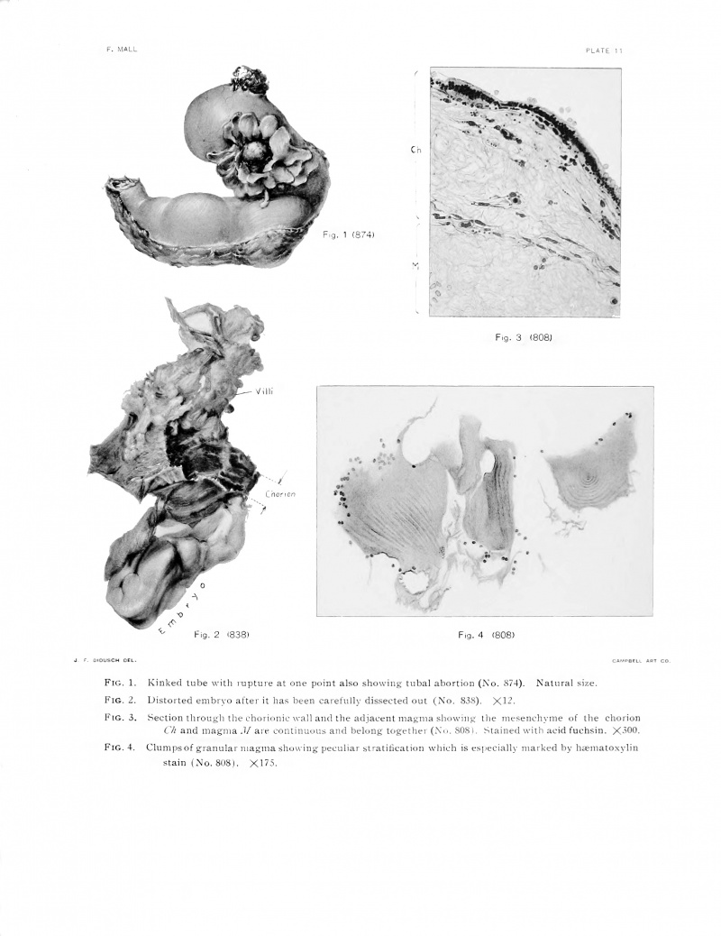
FIG. 1. Kinked tube with rupture at one point also showing tnbal abortion (No. S74). Natural size. FIG. 2. Distorted embryo after it has been carefully dissected out (No. 838). X.12.
FIG. 3. Section through the chorionic wall and the adjacent magma showing UK- iin--eiu'hyme of the cl. C/i and magnia M are continuous and belong together (X<>. 80.S I. Stained with acid fuchsin.
FIG. 4. Clumpsof granular magma showing peculiar stratification which is esi'L-cially marked I vylin
stain (No. 808). XI 75.
| Historic Disclaimer - information about historic embryology pages |
|---|
| Pages where the terms "Historic" (textbooks, papers, people, recommendations) appear on this site, and sections within pages where this disclaimer appears, indicate that the content and scientific understanding are specific to the time of publication. This means that while some scientific descriptions are still accurate, the terminology and interpretation of the developmental mechanisms reflect the understanding at the time of original publication and those of the preceding periods, these terms, interpretations and recommendations may not reflect our current scientific understanding. (More? Embryology History | Historic Embryology Papers) |
Cite this page: Hill, M.A. (2024, April 18) Embryology Book - Contributions to Embryology Carnegie Institution No.1-7. Retrieved from https://embryology.med.unsw.edu.au/embryology/index.php/Book_-_Contributions_to_Embryology_Carnegie_Institution_No.1-7
- © Dr Mark Hill 2024, UNSW Embryology ISBN: 978 0 7334 2609 4 - UNSW CRICOS Provider Code No. 00098G

