Book - Comparative Study of the Sensory Areas of the Human Cortex 1
| Embryology - 20 Apr 2024 |
|---|
| Google Translate - select your language from the list shown below (this will open a new external page) |
|
العربية | català | 中文 | 中國傳統的 | français | Deutsche | עִברִית | हिंदी | bahasa Indonesia | italiano | 日本語 | 한국어 | မြန်မာ | Pilipino | Polskie | português | ਪੰਜਾਬੀ ਦੇ | Română | русский | Español | Swahili | Svensk | ไทย | Türkçe | اردو | ייִדיש | Tiếng Việt These external translations are automated and may not be accurate. (More? About Translations) |
Cajal SR. Comparative Study of the Sensory Areas of the Human Cortex (1899)
- 1899 Human Sensory Cortex: 1. The Visual Cortex | 2. Layer of the Large Stellate Cells | 3. The Sensori-Motor Cortex | Figures | Cajal
| Historic Disclaimer - information about historic embryology pages |
|---|
 |
Lecture I. Visual Cortex
Comparative Study Of The Sensory Areas Of The Human Cortex.
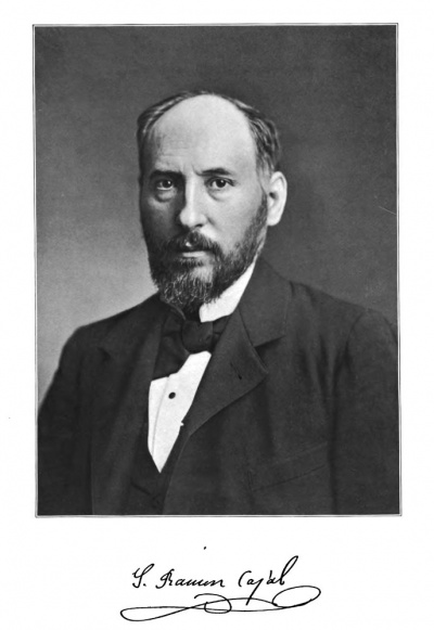
By Santiago Ramón y Cajal
1899

The minute anatomy of the visual cortex (region of the oaloarine fissure, sulcus comu lobulus lingualis) has been already explored by several investigators, among whom we may make particular mention of Meynert, Vicq d'Azyr, Gennari, Krause, Hammarberg, Schlapp, Kolliker, et al. But their very incomplete researches have been performed by such insufficient methods as staining with carmine, the Weigert-Pall method, or that of Nissl with basic anilines — methods which, as is well known, do not suffice at all to demonstrate the total morphology of the elements and the organization of the most delicate nerve plexuses. They led, however, in spite of the difficulties which stood in the way of these first analyrtical attempts, toward a precise differentiation of the visual cortex from other regions of the brain. At the outset two characteristic differences attracted the attention of the first investigators into the structure of the visual cortex: first, the existence of a very thick stratum of granules, subdivided into accessory strata by lamins of molecular appearance; and, second, the presence in the intermediate layers of the cortex of a white lamina formed of meduUated fibres — which lamina may be seen with the unaided eye. This lamina, appearing in cross-section as a white line, has been named, in honor of the writers who first described it, the line of Gennari or Vicq d'Azyr.
For the sake of brevity, we shall omit a detailed description and discussion of the various layers admitted by the authorities on this region ; suffice it to mention in order the eight layers described by Meynert for the human cortex : First, molecular ; the second, layer of small pyramidal cells ; third, layer of nuclei or granules ; fourth, layer of solitary cells ; fifth, layer of intermediate granules ; sixth, layer similar to the fourth, containing nuclei and scattered cells ; seventh, deep nuclear layer ; eighth, layer of fusiform cells. We may also mention the arrangement of layers recently described by Schlapp for the occipital cortex of the monkey : (1) layer of tangential fibres ; (2) layer of external polymorphic cells ; (3) layer of pyramidal cells ; (4) layer of granules ; (5) layer of small solitary cells ; (6) second layer of granules ; (7) layer poor in cells ; (8) layer of internal polymorphic cells.
The investigations which I have made on the human cortex as well as on that of the dog and cat, by both the Nissl and Golgi methods, have led me to distinguish the following layers : —
- Plexiform layer (called molecular layer by authors generally and cell-poor layer by Meynert).
- Layer of small pyramids.
- Layer of medium-sized pyramids.
- Layer of large stellate cells.
- Layer of small stellate cells (called layer of granules by the authors).
- Second plexiform layer, or layer of small pyramidal cells with arched axon.
- Layer of giant pyramidal cells (solitary cells of Meynert).
- Layer of medium sized pyramidal cells with arched ascending axon.
- Layer of fusiform and triangular cells (fusiform cell layer of Meynert).
You see that we have modified current nomenclature by introducing terms which call to mind cellular morphology. For we believe that such trite expressions as "molecular layer," "granular layer," must be banished once for all from scientific language, and they must be replaced by terms which point ont dominant morphological characters in the nerre structures of each layer or some interesting peculiarity relatire to the course and connections of the axis cylinder processes* The number of layers could be easily increased or diminished, because they are not separated by well-marked boundaries, particularly in Nissl*s preparations. Thus the number of layers which I adopt is somewhat arbitrary. By distinguishing, however, nine layers, I have followed a criterion of individualization which seems to me the most convenient and suitable for my exposition of the cortex as a mechanism composed of elements at a certain level which differ in special morphological features from those of neighboring levels. Besides, the number, extent, and size of cells in these layers vary a little in the different median occipital convolutions, as does also the degree of definite nidification, according as we study the convex or concave aspect of the gyri. Our description relates generally to the cortex of the margin of the calcarine fissure, the region where structural differentiation of the visual cortex is most pronounced.
Plexiform Layer
The plexiform or molecular layer is one of the oldest cerebral formations in the phylogenetic series. It presents characters similar to those of the human cortex in all vertebrates except the fishes. This has been fully demonstrated by the researches of comparative histology undertaken by Oyarzun (batrachia), by myself (batrachia, reptilia, and mammalia), by my brother (batrachia, reptilia), by Eddinger (batrachia, reptilia, aves), by CI. Sala (aves). In the visual cortex of man, the structure of this layer coincides perfectly with that which my own researches, as well as those of O. Retzius, have revealed in the motor region. The only modification which may be noted, visible even by Nissl*s method, is its notable thinness in the margins of the calcarine fissure (except in the sulci, and here it appears somewhat thinned). This diminution in thickness, noted by authors generally, depends probably on the small number of medium-sized and giant pyramidal cells in the underlying layers, because it is well known that each pyramidal cell is represented in the plexiform layer by a spray of dendrites. A similar opinion has been expressed by Bevan Lewis in order to explain irregularities in thickness of this layer in different regions of the cortex of the rabbit and guinea-pig. The structure of the plexiform layer is very complex. From my own researches, confirmed largely by those of Retzius, Schafer, KoUiker, and Bevan Lewis, it follows that it consists of an interweaving of the following elements: (a) the radial branches of the small, medium-sized, and giant pyramidal cells, with which we must include in addition those of the so-called polymorphic cells ; (() layer of terminal ramifications of the ascending axons of Martinotti; (c) layer formed by the arborizations of the nerve fibres, terminal or collateral, which come from the white matter ; ((2) layer of special or horizontal cells of the first layer (Cajal's cells, of Retzius) ; (e) layer of small and medium-sized stellate cells with short axons ; (/) layer of neuroglia cells, well described by Martinotti, Retzius, and Andriesen.
a. Terminal Arborizations of the Pyramidal Cells (Fig. 4). — As my observations have shown in case of the mammalian cortex, and those of Retzius for the human foetus, the radial trunk of the pyramidal cells does not end, as Golgi and Martinotti supposed, in a point entwined by neuroglia elements in connection with the blood-vessels, but in a spray of varicose dendrites covered with contact granules, spreading out sometimes over a considerable area of the plexiform layer. In my first work on the cerebral cortex, I thought that the only cells whose terminal dendrites reached up to the first layer were the medium-sized, small, and giant pyramidal cells; but my latest researches have enabled me to discover that all cells possessing a radial stem, without exception, including even those of the deeper layers, are represented in the plexiform layer by a terminal dendritic arborization. It is without doubt an important structural law whose physiological import must be very considerable. We may observe that large trunks which arise from the giant pyramids divide into a spray with very long and thick branches having their distribution in the deeper level, while the slender stems emanating from the medium and small sized pyramids form an arborization of numerous slender branches of limited extension and distributed particularly through the superficial laminae of the plexiform layer. This distribution, which is not absolutely constant, leads us to surmise that the terminal arborizations of each kind of pyramidal cell come into contact with special neuritic terminal arborizations in traversing this first layer.
The trunk and end brush intended for the first layer appear not only in preparations made by the chromate of silver method; for I have stained them perfectly with methylene blue (method of Ehrlich-Bethe) in case of young animals, and also in adult gyrencephalous mammals, such as the dog and cat. Besides, in good preparations by Ehrlich*s method, particularly when fixation has been made a short time after the impregnation, one may see very distinctly the contact granules of the dendrites, processes which I was first to describe and whose existence has been confirmed by many investigators since. With methylene blue they present the same appearance as in Golgi preparations, i.e. they are slender and short, stand out at a right angle, are sometimes divided, and end freely in a rounded knob. This proves, accordingly, how groundless are all the gratuitous objections which have been brought against the preexistence of these appendages, as well as against their mode of termination. Among the entirely arbitrary conjectures which have been made as to the disposition of these appendages we include also W. Hill*s opinion, who considers them the fibres of a reticulum that is incompletely stained by means of the chromate of silver. We must proclaim emphatically that at present there is no method of staining cellular processes that is capable of disproving the agreeing results of the methods of Golgi, Ehrlich, and Cox. Whoever, having as a foundation the revelations of any one of these methods, has considered it possible to demonstrate the existence of such a reticulum has only exposed to view his own lack of experience in handling these important means of analysis.
b. Special or Horliontal Cells of the Plexiform layer. — These interesting elements, which I discovered in the cortices of the small mammals (rat, rabbit, guinea-pig), have been successfully investigated by Retzius in case of man, as well as by my brother in batrachians and reptiles, and by Veratti in the rabbit's embryo. They present in the visual cortex, where I have stained them very often, the same characters as in other regions of the brain. As I have already described these elements elsewhere, I shall give here only an outline, to which I may add a few remarks derived from my recent observations upon man (Fig. 2).

Fig. 2
Following the example of Retzius, when we study the horizontal cells by Golgi*s method in a human fcetus from the seventh to the ninth month, or in case of a newborn babe, we notice that they are distributed throughout the entire thickness of the plexiform layer, but are especially numerous in close proximity to th6 pia. Their form is very variable, sometimes fusiform or triangular, and again stellate, with the angles extending out into the long processes. But the characteristic feature of these elements is due to the fact that their processes, which are variable in number and yery large at their origin, give rise, after a few divisional to an extraordinary number of yariooee horizontal fibres, extremely long, from whioh spring at ri^t angles numberless ascending secondary branches terminating in rounded knobs near the cerebral surface* Very often the superior surface of the cell body also gires rise to some of these ascending branches, which sometimes have a considerable thickness.
In what way do these tangential fibres terminate? Is it possible to diMem among them certain processes possessing the characters of axons?
Upon careful examination of the best preparations obtained from cortices of human embryos, we discover easily that these processes, when they become very fine, have all the appearances peculiar to axons. There is no morphological distinction which would enable us to distinguish the two classes or species of cellular processes. That which most strikes one is the enormous length of their horizontal fibres (tangential fibres of Retzius). One can follow them for two or three tenths of a millimeter without being able to discover their true termination. However, in certain cases it is possible to demonstrate that the tangential fibres, after having given rise to a great number d vertical twigs, become thinner and finer, and finally subdivide into terminal branchlets, which spread out under the pia or in the superficial lamins of the first layer.
On comparing these cells of the human brain with their homologues in tiie higher mammals (rabbit, cat, etc), we diicover that among the latter they give rise to a relatively small number of tangential branches, and that these extend a much shorter distance. This is the reason we consider the remarkable profusion and the extreme length of t&e horizontal fibres as one at the most characteristic features of the human cortex.
Retrius did not succeed in staining the horizontal cells in man except in the foetal period. Accordingly, it was impossible to know what becomes of these elements in the adult, and whether, as Retzius is inclined to think, all the processes that we find in the embryonic period persist My recent researches on the cortex of infants fifteen months and even fifteen and twenty days old, in which I have been successful in staining the horizontal cells, suffice to furnish a few data which, if they do not solve the problem once for all, at any rate place the question in a somewhat more fiivorable light.
When we examine the plexiform layer of a babe fifteen days old,, we find considerable changes in the horizontal cells. First of all we notice that they have become smaller, and that the tangential processes have diminished in diameter while they have become notably lengthened. But the peculiarity which most strikes the attention is the almost total disappearance of the ascending collateral branches. This atrophy begins in a progressive thinning of the processes and in the reabsorption of their terminal varicosities ; then the whole branch disappears, so that the only structures left are the horizontal fibres, whose ensemble forms throughout the thickness of the plexif orm layer a system of parallel fibres of enormous length. There are places, however, where the ascending branches persist, but very much changed as to their direction, having become oblique instead of vertical, becoming branched several times, and terminating in the plexiform layer without reaching so far up toward the pia as before. In a word, most of the vertical branches seem to me to represent an embryonic arrangement corresponding to the interstices, for the most part vertical, between the epithelial cells of the cerebral cortex of the fcBtus, which proves once more, as I have demonstrated in other nerve centres, that during the period of evolution the neuron is the locus of a double series of functions: on the one side a vegetative building up of the dendrites ; on the other, reabsorptions and transformations of the cells which persist.
Have the horizontal cells with which we are now concerned a true functional process ? In case this is so, what is the part played by these elements in the vast system of nervous relations established in the plexiform layer ?
In preparations of the human brain stained with chromate of silver, it must be confessed, it is not easy to solve this important question, since the purely morphologfical criterion, which is sufficient to distinguish the axon in other neurons, cannot be applied to horizontal cells, all the processes of which, on becoming finer, have the form of true axons. Thus, in spite of Veratti's affirmation, we believe that this method will shed no light upon the subject, even when applied to embryos. In order to approximate to any solution of the problem, we must use a method capable of staining nerve prolongations in a manner to differentiate them from dendrites. It was only after using Ehrlich's methylene-blue method upon the motor and visual cortex of the cat that I became convinced that the horizontal cells have in reality a very long axon, which is provided with a .medullary sheath. The other processes, which we have called horizontal fibres, represent true dendrites, as is shown by two peculiarities: the great hcHity with which they take methylene blue, and their pronounced varicosity after fixation with ammonium molybdate. We must repeat that this varicose alteration, which is a striking modification in the form of cellular prolongations, presents itself only in dendrites. The neurites maintain perfectly, with methylene blue, their normal contours, unless exposure to the air, necessary to obtain the selective staining, has been too long.
As to the axon, it may be sufficiently well demonstrated in horizontal sections of the plexiform layer in the form of a pale blue fibre, except the initial portion and the nodes, which present a dark blue staining. This is a property of all parts of a fibre not surrounded with a medullary sheath. At the point of certain constrictions we may succeed in discovering a few collaterals springing out at right angles, provided also with myeline sheaths. Finally, one is sometimes so fortunate as to discover in an axon of this kind true bifurcations situated at a great distance from the cell of origin, but always in the plane of the plexiform layer. Unfortunately, the methylene blue does not stain the terminal nerve arborizations. This has prevented me from learning in just what way these axons terminate and with what axons they are dynamically associated. It is possible that certain heavy horizontal fibres come into contact with the horizontal cells, since they never bend downward toward the underlying layers, as do the medium-sized and finest medullated fibres. They belong probably to the terminal arborizations of Martinotti^s ascending axons and, perhaps, also to the collaterals and terminals coming in from the white matter.
e. Cells with a Short Axon (Fig. 8, (7, H.F). — A few years ago, while studying the cerebral cortex of the small mammals, I discovered, besides the gigantic horizontal cells, other elements which I called polygonal cells. These are characterized by their stellate form and by their short axon, which ramifies and ends within the limits of the plexiform layer. These cells, whose existence neither Schaf er nor Lewis seem to have been able to confirm, — no doubt on account of the insufficiency of their attempts to obtain an impregnation of them, — are much more abundant than might have been supposed from my first observations. However, I must acknowledge that, they are not at all easily impregnated with chromate of silver and that, in order to find a sufficient number for study, we must make a great many attempts at staining them. On the other hand, Ehrlich*s method stains them very readily in the dog and rabbit. In these animals — and I think that it holds true also in man — the plexiform layer of the cerebrum is as richly supplied with elements with a short axon as the molecular layer of the cerebellar cortex. They occur in all levels of the layer and differ remarkably in size and shape. The majority of them are stellate and are comparable in size to other cells with short axons that occur in the deeper layers of the cortex. Others are smaller, resembling in their minuteness the g^ranules of the cerebellum. But whether large or smaU, the morphological characters of these elements are very similar. Their dendrites are divergent, extremely branched, and distributed exclusively to the plexiform layer* Their neurites are Qgoftlly very short, subdiride in a most complicated manner in the neighborhood of the cell, but never crota the deep boundary of the first layer.

Fig. 3. — Cells and neuritic terminal arborisations in the 1st and 2d layers ; visual cortex of infant 20 days old. A and B, neuritic plexus, extremely fine and dense, situated in the layer of small pyramids ; C, an analogous arborization, but not so dense ; D, a small cell whose ascending axon forms a similar arborization ; E, spider-shaped stellate cell of the 1st layer ; F, G, cells with short axon branching loosely in the plexiform layer ; a, axon.
From the point of view of the direction and length of their neurites all these elements may be classified into three varieties : (1) Stellar cells with horizontal neurite which becomes resolved after a varying distance, generally very long, into a terminal arborization which has the appearance of being connected with the terminal branches of the remote pyramids. (2) Cells of generally smaller size whose neurite branches either laterally or vertically from the cell body, but always at a moderate distance (Fig. 8, <?, J^. (8) Very small cells (which I discovered recently in the human cerebral cortex) provided with numerous fine, divergent, and slightly branched dendrites, whose neurite, extremely slender, breaks up near its origin into a dense arborization, exceedingly fine and complicated We shall designate these elementa dwarf or spider-shaped cells. They may be found, as we shall see, in all the layers of the cortex (Fig 8, ^) To sum up : bearing in mind the form of cell bodies and formation and connection of axons, all the stellate cells of the plexiform layer, including the horizontal or special cells, seem to me similar to the stellate cells of the molecular layer of the cerebellum and to those which occur in the layers of the same name in the eormn ammonU and fascia deniaia. Their function is probably to establish connections between terminal arborizations as yet imperfectly made out, possibly those formed by the ascending axons of Martinotti, or the association fibres coming up from the white matter with the terminal branches of the pyramidal cells. The function of the great horizontal cells would seem to be to establish connections between elements, that is to say between terminal neuritic arborizations and radial dendrites, separated by very considerable distances; while the medium-sized and small elements, with their short axons, would perform the same associative function at short or moderate distances.
d. Hartinottl's AtcsmUng Fibrea. — There is no Uck of these in the visual cortex, although it has seemed to me that they are not so numerous as in other regions of the brain. Their terminal ramifications, well known from the researches of Martinotti as well as my own, occupy really the whole plexiform layer, where they extend over wide areas, distributing themselves preferably into its deeper levels and coming in contact with cells with short axons and, possibly, also with the large horizontal cells.
Granting that the cells of origin for these fibres lie in layers of the cortex that contain sensory fibres, we might suppose that Martinotti's ascending axons represent intermediate links placed vertically between these sensory fibres and cells with short axon in the plexiform layer. And as these are connected, perhaps, with the dendrites of the pyramidal cells, the result would be that the sensory stimuli, entering the cortex in this indirect way, would be compelled to traverse two intercalated nerve cells before reaching the pyramids.
6. Neuroglia Cells. — These conform in the visual cortex to the wellknown types of other cerebral regions. We find accordingly : (1) Cells with long radii, the marginal cells well described by Martinotti, which lie just under the pia. They emit long, smooth, descending processes radiating across the plexiform layer, ending at different levels both of this and of the layer of small pyramids; (2) Cells with short radii. These elements, long since described by Golgi, and described in detail by Retzius, by myself, Andriesen, Kolliker, and others, are characterized by their form, very often stellate or fusiform, by their location in all levels of the plexiform layer, and by the great number of their processes, short, spongy, branching, and bristling with innumerable contact granules, which penetrate into the spaces lying between the neuro-protoplasmic plexus and are well spread over the interstices of the elements which must not come into contact. It is in virtue of this intricate relation between these appendages and the cell bodies and dendrites, as well as for other reasons which we have not time to dilate upon here, that we attribute to the neuroglia elements with short processes an insulating role. According to my view, they prevent inopportune contacts, while their processes exercise due regard to all points of cells or fibres where contacts exist and nerve currents pass.
Layer of Small Pyramids
This layer is well separated from the 1st, but blends by insensible gradations with the 3rd, or layer of medium-sized pyramidal cells (Fig. 4,5).
Examined in.Nissl preparations this layer presents a great number of small pyramids, very poor in chromatic granules and separated by a plexus of fibrils much more dense than in the case of cells of the deeper layers. We find also, scattered irregularly, stellate or triangular cells larger than the pyramids. Theee are the giant cells with short axon, as is shown in good ohromate of silver preparations (Fig. 5, 2>, CT). We shall now discoss the cells of this layer, beginning with the pyramids. Pyramids. — The morphology and relations of these cells being well known since the researches of Golgi, Retrius, and myself, I shall limit my remarks to a bare mention of a few peculiarities of their disposition in the visual cortex.
Fig. 4. — Saudi and madimn-daad otUi of thm yfrnud oortas of ab lafuii ao days old (oatoarias i). A, Plazifdna layer ; B, layar of amall pyrramkU ; C, layar of madliiaKtlaad pyianlds; «, azoB ; b, rtearreot eoUaUral ; e, dandritic tnmk of i^aat pyraaidd.
It will be noticed that these cells are generally smaller and more numerous in the visual centres than in other cortical areas. Sometimes the more superficial cells are arranged in one or two regular files and separated from those beneath by a fine dense plexus of fibres.
The small pyramids give rise to the following processes : an axial dendrite, often bifurcated near its origin, which runs to the plexif orm layer and terminates in a spray of fine branches, which often ascend to the neighborhood of the pia; basilar divergent dendrites, rather long and repeatedly branched; and, finally, a fine descending axon, which, in most favorable specimens, can be followed down to the neighborhood of the white matter. From the initial portion of its course spring three, four, or a larger number of collateral processes, which traverse, with many subdivisions, in a horizontal or oblique direction, a very considerable extent of the second layer. From the small pyramids lying close to the plexif orm layer, and even from some cells more deeply situated, the first two collaterals recurve, ascending sometimes, as Schafer has discovered, up to their termination in the first layer. However, this termination in the first layer is much less frequent than might be inferred from this authority's descriptions and drawings. In our preparations of the visual and motor cortex of a child a few days old and of a cat twenty-five days old, the great majority of the recurrent collaterals do not cross the boundary of the second layer. Here, in conjunction with many neurites belonging to cells with short axons, they assist in forming a very dense plexus, which contains in its meshes the primary dendrites of the small pyramids. Grenerally, — and this may be considered as an answer to the authorities who strive to convert the recurrent course of the collaterals into an argument for the doctrine of the cellulipetal conduction of these fibres (v. Lenhossek, Schafer), — I may affirm that the vast majority of the initial neuritic collaterals — and I consider such all those that arise within the gray matter — always come into contact with some of the dendrites belonging to homologous nerve cells situated at different levels of the same cortical formation. When the cells to which they correspond lie in the same or a deeper plane, the collaterals intended for th^u take a horizontal, descending, or oblique course ; but if the cells of the same category are situated in a more superficial plane than the point of origin of the collateral, they must describe a recurrent arc in order to reach their destination.
Layer of Medium-sized Pyramids
Being a continaatdon by insensible gradations of the small pyramidal layer, it contains cells of precisely similar form, differing from the cells of the second layer only in their some^diat greater size, their longer radial dendrite, and, ordinarily, by a larger number of neoritic collaterals (Fig. 4, (7). In the deeper level of this layer may be observed — very seldom, however — large pyramidal cells, but not so large as those situated in the seventh layer.
Cells with Short Axon of the Second and Third Layers. — These elements, almost as numerous as the pyramidal cells themselves, may be seen scattered irregularly throughout the entire thickness of the two layers. They are generally more numerous near the limits of these layers, that is to say, in the superficial portion of the second and in the deeper level of the third layer.
Although in form and size these elements are very variable, and although there are transitional forms which make it often difficult to distinguish between them and to subdivide them into well-pronounced types, still, by considering the size of the cell body and the character of the axon, they may be divided into the following five classes : (a) cells with short ascending axon ; (6) cells with short descending axon ; (0) celb with horizontal or oUique axon ; (d) dwarf or spider-shaped elements ; (e) fusiform or bipanicled cells, whose axon breaks up into a fibrillar arborization.
a. CeU$ with Aneending Axon (Fig. 5, a, B). — As may be seen in Fig. 5, these cells belong to two principal varieties : (a) Gigantic cells, with long dendrites (Fig. 5, A, (7). These are quite numerous in the visual cortex, where they occupy preferably the deep portion of the third layer. Their form is stellate, sometimes fusiform or triangular. From their angles arise several varicose, thick, and very long dendrites, often disposed as two brushes, the one ascending, the other desoending. The axon takes its origin either from the cell body or from a dendrite. Sometimes it describes an arc, whose concavity is toward the surface, on its way outward to become resolved into an arborization of very few branches. The characteristic feature of this arborization is the enormous length and the horizontal or oblique direction of its terminal twigs. These traverse a very considerable portion of the second and third layers, where they make contact with numberless pyrramidal cells. It may be added that theee gigantic cells may be recognized even in Niasl preparations by their stellate form and considerable size. They correspond, probably, to the globular cells of Bevan Lewis and other writers. (&) Medium-sized type : This is a fusiform or stellate cell, whose size does not exceed that of the small or medium-sized pyramids. It is characterized above all by its axon, which is slender and ascending, and which terminates in a complicated arborization with many varicose branches and with relatively small spread at varying levels of the second and third layers. As to the dendrites, they appear varicose and diverge in all directions, but usually do not extend to the first layer (Fig. 5, Fy and Fig. 8, 2>).
Fig. 6. Large stellate cells haying short ascending axons, 2d and 3d layers, yisoal cortex, infant 10 days old. A, elements of the 3d layer with axons divided into long horizontal branches ; B, small oell with arched axon from the layer of small pyramids ; C, large cell with arched axon ; 2), large cell from the boundary of the Ist layer ; F^ cell with arched ascending axon branching in a most complicated manner; a^a^a^ axons.
&. Cells with Descending Axons. — These are stellate, triangular, or fusiform, of medium size, and provided with many ascending and descending dendrites. They occur chiefly, as has been pointed out by Schafer for other regions of the cortex, along the superficial boundary of the layer of small pyramids (Fig. 5, £, and 6, (7). Their axons descend through the second and sometimes through the third layer, giving off to them a few collaterals, and terminate in a diffuse arborization throughout the different levels of these layers. Very frequently this axon, after descending a certain distance, emitting a few collaterals to the layer of small or medium-sized pyramids, traces an arc with concavity toward the surface and ascends to terminate in an arborization, very complicated and with exceedingly varicose branches, in the layer of small pyramids close to the plexiform layer (Fig. 5, £). As seen in Fig. 6, which reproduces certain cells of short axons from the visual cortex of the cat, these elements with descending axons are very numerous in other gyrencephalous mammals. We also find a variety of cell, recognized in man, pyriform, uni-polar, whose single descending process gives rise to a bouquet of varicose dendrites and an axon (Fig. 6, a, &). The collaterals and terminal arborizations of these axons form in the cat a dense plexus throughout the superficial plane of the layer of small pyramids.
The great number of cells with short axons which occur in the most superficial lamina of the layer of small pyramids has induced certain writers, such as Schafer and Schlapp, to consider this transitional region as a special layer, which they call the layer of superficial polymorphic cells. We cannot subscribe to this innovation because, in spite of the great number of these cells, this transitional lamina contains also a large number of small pyramids, that is to say, cells which, in addition to their morphologioal varieties, have the same oonnectioiis as ordinary pyramidal cells. Of course, if for the subdivision of the cortex into layers we take as our basis of classification the form of cell bodies, independently of other characters, we might be entitled to differentiate between the first and second layer consisting chiefly of stellate cells ; because in this region, as is well known, the small pyramids have a stellate or triangular form. But, in assigning to an element a place in his classification, one must not decide from the form alone, which in case of superficially placed pyramids is a function of their position. In fact, we find that the form of these cells varies according to their proximity to the plexiform layer. The true characteristic of a pyramidal cell consists in the presence of a long axon extending down to the white matter and of a spray of dendrites (supported or not by an intermediate trunk) spreading up into the plexiform layer. Now, in the light of such a criterion, it is easy to see that sufficient reason does not exist for making out of the most sui)erficial pyramids a distinct category of cells to be used as a basis for the creation of a new cortical layer.
Fig. 6.— Cells with short axons from the layer of small pyramids, yisnal cortex of cat aged 28 days, a, 6, small psrriform cells with short descending axons ; c, cell with arched axon ; e, /, cells with descending axons distributed to the medinm-sized pyramids of 3d layer.
c. cells with Horizontal or Oblique Axon (Fig. 7). — These elements, which are angular or fusiform, with their long axes more or less horizontal, possess few, but rather long, dendrites. Their axon arises generally from the lateral aspect of the cell body or from a thick polar dendrite, takes from the first a horizontal or oblique direction and, after giving off a few collaterals, terminates, sometimes after extending to a considerable distance, in an arborization widely spread but with few branches. In certain cells of this category, it is shorter and subdivides in the immediate neighborhood of the cell body (Fig. 7, JST, CT).
d. Dwarf or Spider^haped Cells. — Brought to our attention by CI. Sala in the corpus striatum of birds, mentioned also by my brother in the cerebral cortex of batrachians and reptiles, these strange elements are notably abundant and of very pronounced character in the cerebral cortex of man and gyrencephalous mammals. They are found irregularly scattered in all layers of the visual area. Their soma is very small, not exceeding the diameter of the nucleus by more than five or six /i. About the nucleus is a thin lamina of protoplasm which is drawn out into a great number of dendrites, delicately varicose, radiating, slightly branched and short. The appearance of these dendrites is such tliat one might mistake the cell, at first sight, for a neuroglia corpuscle with short processes. But, examining them with a high power, we recognize at once that their slender dendrites do not possess collateral appendages (contact granules), so characteristic of processes of neuroglia cells. F^inally, attentive examination reveals the axon, a delicate fibre, which becomea resolved immediately into a very dense varicose arborization of incompar^ able fineness. Often this terminal plexus is so extremely fine that it appears through an ordinary objective as a yellowish or brownish spot in the neighborhood of the cell and resembling somewhat a granular precipitate. In some cases this arborization is coarser and can be seen with a Zeiss objective D or E. At the level of the superior boundary of the layer of small pyramids, in the visual cortex of the child and even of other mammals, may often be seen a dense plexus of exceedingly slender branching fibrils. Their original fibre appears to come from the deeper levels of the 2d layer (Fig. 8, A^ B^ CT). These terminal plexuses often take the impregnation irregolarlyt which gives the appearance of brownish or coffee-colored spots scattered and sometimes arranged in a row just underneath the plexiform layer. At first I was not successful in tracking satisfactorily the fibres of origin and, therefore, hesitated as to stating the significance of these interesting arborizations. Very recently, however, in two or three fortunate specimens I have been able to demonstrate the connection between this plexus and the fine ascending axons of certain small cells situated in the deeper level of the 2d or outer level of the 8d layer. I am, therefore, now inclined to consider this intermediate, or subplexiform, nerve plexus as consisting of terminal arborizations intended for the small pyramids. The fibres of origin spring from more deeply situated spider-shaped cells very hard to impregnate. I may add that these plexuses are not lacking in the oat and dog, although in these ^wimaU the fibrillsB are not so numerous nor so extremely fine as in the human brain. Permit me also to add that they occur in all regions of the cortex, although up to the present we have obtained the best impregnation of them in the visual area.
Fio. 7. — Cells with short horizontal or oblique axons situated in the 2d and 3d layers, Yisnal cortex of infant a few days old. A^ B, cells with axons almost horizontal from 2d layer ; C, D, E, cells with short axon diffusely branched ; F, H, I, pyriform cells of the 1st layer, whose significance is still uncertain; O, small cell with yery short axon diffusely branching within the 1st layer.
e. SmaU BipanieUd 0M$. — In the visual region, as well as in other areas, of the human cortex we find in profusion certain small cells vertically elongated. Their axon presents the very singular feature of breaking up into long slender brushes of terminal fibrills. At first, I thought that these singular cells were forms characteristic of the acoustic area, for here they are remarkably developed and very numerous. Further investigation, however, has convinced me that they occur in all parts of the cortex, disposed in greatest numbers along the lower level of the 2d and 8d layers (Fig. 8 and Fig. 11, Jf, F).
As stated above, we are discussing the small spindle-shaped cells with poles radially disposed, which give rise to groups of dendrites, slender, unprovided with contact granules, very finely varicose, and often arranged in long ascending and descending brushes. In some cases these are so fine that on superficial examination they might be mistaken for delicate neuritic arborizations. But the most striking peculiarity of these cells concerns the subdivisions and course of their axons. This process is very delicate. It ascends or descends a certain distance, then generally gives off a few collaterals at right angles which soon subdivide into ascending or descending fibrillse, and finally it breaks up into brushes of very slender filaments which run radially, extending throughout almost the entire thickness of the cortex. As a whole this arborization with its initial collaterals forms one or several parallel brushes, the fibrils of which skirt the trunks of the pyramids and adapt themselves to the cell bodies, over which they appear to creep, like the creeping fibres of the cerebellum on the branches and bodies of the Purkinje cells.
Fig. 8. — Small fusiform, bipanided cella from anditory cortex of infant (let temporal •oonyolation). A, cell giving origin to a descending axon moderately branched ; B, cell whose axon breaks ap into a number of pencils of yery long ascending and descending fibrils ; a, axon. (Examined with Zeiss apo<chromatic obj. 1.30.)
In the brain of the human infant at birth these arborizations have not attained complete development and present but few vertical branchlets. It is not until twenty or thirty days after birth that we can observe the long and complicated terminal brushes. In certain areas, the acoustic, for example, each neurite may form as many as five ascending or descending brushes. The fibrils of which they consist are so delicate that in order to see them well we must use the highest apochromatic objectives.
If now we consider all the different kinds of cells having short axons, of which we have given a somewhat fastidious description, from the point of view of their connections and their probable functions, we may characterize them as special cells of association. The form of their cell body and the disposition of the axon vary according to the number, form, and position of the cells to which they must convey nerve stimali. Thos cells with a horizontal axon most be intended to transmit impulses to elements, probably pyramidal cells, which occur at the same level in the cortex. Those whose axon is vertical, ascending or descending, would naturally transmit impulses to elements of different layers. Those which are bipanicled would serve to associate dynamically a great number of pyramids in vertical series. Finally, the small, spider-shaped cells may have for their function association of groups of pyramids very close together. Unfortunately for this theory, we do not know from which nerve fibres all these elements of association receive their initial stimuli. Accordingly, we must be resigned to remain in ignorance as to the path of the afferent impulses and, as well, in regard to the special influence which these elements must exercise. It seems very probable, however, that their function consists not only in facilitating the spread of incoming stimuli, but also in adding to it something new, some specific modification which cannot now be determined. We shall return to this point in our general conclusions upon this work. But we may see from the above how many paths nature has opened up to render association of nerve impulses possible in every direction and through any distance. That which proves the importance of these association cells and leads us to surmise that they play an important psychic r61e is the fact that they are extremely numerous in the human brain. They are found in the animal brain as well, but are not numerous and are usually confined to the boundary of the 1st layer.
I conclude here my exposition of the prosy topics that I chose as the theme of this lecture. And nothing remains except to thank you for the attention and good will which you have shown me in spite of the extreme dryness of the subject-matter.
- 1899 Human Sensory Cortex: 1. The Visual Cortex | 2. Layer of the Large Stellate Cells | 3. The Sensori-Motor Cortex | Figures | Cajal
| Historic Disclaimer - information about historic embryology pages |
|---|
|
Glossary Links
- Glossary: A | B | C | D | E | F | G | H | I | J | K | L | M | N | O | P | Q | R | S | T | U | V | W | X | Y | Z | Numbers | Symbols | Term Link
Cite this page: Hill, M.A. (2024, April 20) Embryology Book - Comparative Study of the Sensory Areas of the Human Cortex 1. Retrieved from https://embryology.med.unsw.edu.au/embryology/index.php/Book_-_Comparative_Study_of_the_Sensory_Areas_of_the_Human_Cortex_1
- © Dr Mark Hill 2024, UNSW Embryology ISBN: 978 0 7334 2609 4 - UNSW CRICOS Provider Code No. 00098G