Template:Lisser1911 figure gallery
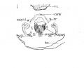
Fig 1 Cross section of human Embryo no. 109 (10.5.) to show cricoid cartilage and M. criooarytaenoideus posterior.
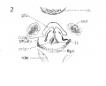
Fig 2 Cross section of human Embryo no. 109 (10.5mm.) to show thyreoid cartilage, M. criooarytaenoideus lateralis,
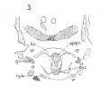
Fig 3 Frontal section of human Embryo no. 317 (12.5 mm.) to show epiglottis thyreoid cartilage, and hyoid bone. Ant. Card., anterior cardinal vein.
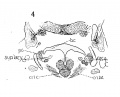
Fig 4 Frontal section of human Embryo no. 317 (12.5 mm.) to show superior laryngeal nerve, sup. /ary., superior laryngeal nerve; car. a., carotid artery.
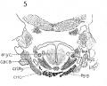
Fig 5 Frontal section of human Embryo no. 317(12.5 mm.) to show M. crico-thyreoideus and arytaenoid masses.
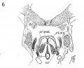
Fig 6 Frontal section of human Embryo no. 317 (12.5 mm.) to show M. cricoary taenoideus posterior, interarytaenoideus and nerve recurrens.
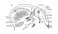
Fig 7 Sagittal section of human Embryo no. 144 (14 mm.) to show, especially, M. interarytaenoideus.
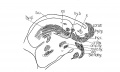
Fig 8 Sagittal section of human Embryo no. 144(14 mm.) to show hyoid bone and thyreoid cartilage, M. cricothyreoideus, and tongue region
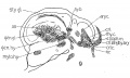
Fig 9 Sagittal section of human Embryo no. 144 (14 mm.)
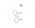
Fig 10 Graphic reconstruction of cricoid and arytaenoid cartilages in human Embryo no. 43 (16 mm.).
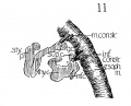
Fig 11 Graphic reconstructions of pharyngeal constrictors in human Embryo no. 43 (16 mm.)
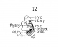
Fig 12 Graphic reconstruction of laryngeal musculature in human Embryo no. 43 (16 mm.).
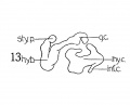
Fig 13 Graphic reconstruction of thyreoid cartilage hyoid bone, and styloid process in human Embryo no. 43 (16 ram.).
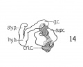
Fig 14 Graphic reconstructions of larynx cartilages in human Embryo no. 43 (16 mm.).
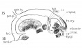
Fig. 15 Sagittal section of human Embryo no. 43 (16 mm.) to show laryngeal musculature and nerve recurrens. Meek, c, Meckel's cartilage.
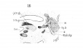
Fig. 16 Sagittal section of human Embryo no. 43 (16 mm.) laryngeal region, s. m. gl., submaxillary gland.
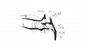
Fig. 17 Graphic reconstruction of 9th, 10th, and 12th cranial nerves in larynx region of human Embryo no. 43 (16 mm.), ana., anastomosis between superior laryngeal and inferior laryngeal nerves; m br., motor branch of superior laryngeal nerve.
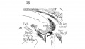
Fig. 18 Sagittal section of human Embryo no. 43 (16 mm.) laryngeal region.
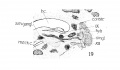
Fig. 19 Sagittal section of human Embryo no. 43 (16 mm.) laryngeal region, s. m. gangl., submaxillary ganglion.
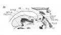
Fig. 20 Sagittal section of human Embryo no. 43 (16 mm.) laryngeal region.
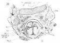
Fig. 21 Frontal section of human Embryo no. 128 (19.5 mm.) to show thyreoid cartihige and M. cricoarytaenoideus lateralis, occ. h., occipital bone.
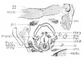
Fig. 22 Frontal section of human Embryo no. 128 (19.5 mm.) laryngeal muscles and cartilages.
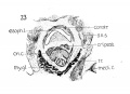
Fig. 23 Frontal section of human Embryo no. 128 (19.5 mm.) to show cricoid cartilage, M. cricoarytaenoideus posterior, and thyreoid gland.
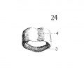
Fig. 24 Graphic reconstruction of cricoid cartilage, posterior veiw, in human Embryo no. 22 (20 mm.) 3, anterior arch; J!^, posterior arch.
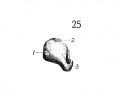
Fig. 25 Graphic reconstruction of cricoid cartilage, lateral veiw in human Embryo no. 22 (20 mm.). /, articular facet for thyreoid cartilage; 2, articular facet for arytaenoid cartilage.
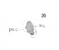
Fig. 26 Embryo no. 22 same as fig. 25, showing extent of choudrification. tr. c, true cartilage; pre. c, pre-cartilage (condensed mesenchyma).
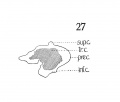
Fig. 27 Graphic reconstruction thyreoid cartilage in human Embryo no. 22(20 mm.) showing extent of chodrification.
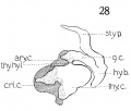
Fig. 28 Graphic reconstruction of laryngeal cartilages in human Embryo no. 22 20 mm.) thy. hy., 1. thyreohyoid ligament.
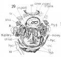
Fig. 29 Cross section of human Embryo no. 22 (20 mm.) to show especially superior laryngeal nerve and nerve recurrens.
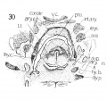
Fig. 30 Cross section of human Embryo no. 22 (20 mm.) to show, especially, M. interarytaenoideus and aryepiglotticus. This section shows at the mark (x) the tendency to continuity between the laryngeal and pharj'ngeal musculature, as - mentioned by Strazza.
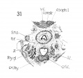
Fig. 31 Cross section of human Embryo no. 22 (20 mm.) (very low in laryngeal region).
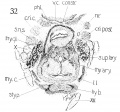
Fig. 32 Cross section of human Embryo no. 22 (20 mm. ) to show thyreoid cartilage, cricoid cartilage, and hyoid bone. Also M's cricoarytaenoideus posterior and thyreorarytaenoideus.
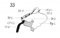
Fig. 33 Graphic reconstruction of nerve recurrens and its branches in relation to the laryngeal muscles and cartilages in humanEmbryo no. 22
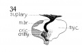
Fig. 34 Graphic reconstruction of motor branch of superior laryngeal nerve in human Embryo no. 22 (20 mm)
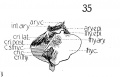
Fig. 35 Graphic reconstructions of laryngeal muscles, and cartilages, and their relations in human Embryo no. 22 (20 mm.) ary. epi., M. aryepiglotticus; thy. epi., M. thyreoepiglotticus; c. s., thy. c, cut surface thyreoid cartilage.
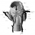
Fig. 36 Wax model of laryngeal muscles and nerves in human Embryo no. 22
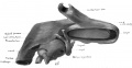
Fig. 37 Wax model of laryngeal region in human Embryo no. 22(20 mm.). (seen from below)
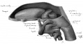
Fig. 38 Wax model of laryngeal region in human Embryo no. 22 (20 mm.). (drawn from the left side)
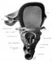
Fig. 39 Wax model of laryngeal region in human Embryo no. 22 (20 mm.). (drawn from below)


