Uploads by MarkHill
From Embryology
This special page shows all uploaded files.
| Date | Name | Thumbnail | Size | Description | Versions |
|---|---|---|---|---|---|
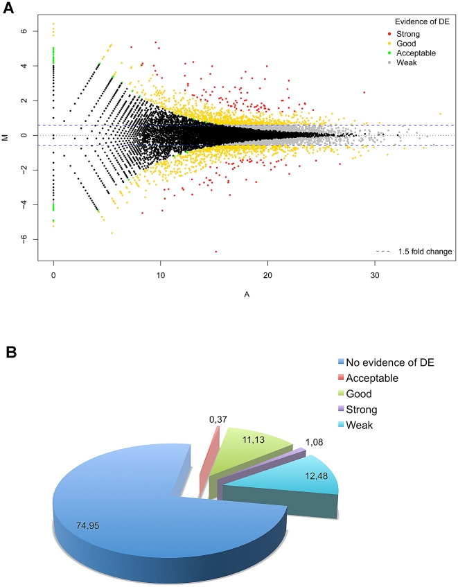
| 18:33, 11 August 2011 | Differentially expressed RefSeq genes in human trisomy 21.jpg (file) |  |
171 KB | Reverted to version as of 02:46, 11 August 2011 | 3 |
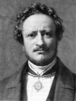
| 12:29, 20 June 2011 | Johannes Muller.jpg (file) |  |
20 KB | ==Johannes Müller (1801-1858)== Category:Historic Embryology | 1 |
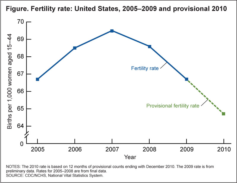
| 11:00, 20 June 2011 | USA births fertility 2010.jpg (file) |  |
54 KB | ==USA Births and Fertility 2010== '''Recent Trends in Births and Fertility Rates Through 2010'''<ref>Sutton PD, Hamilton BE, Mathews TJ. '''Recent Trends in Births and Fertility Rates Through 2010''' NCHS data brief MD: National Center for Health Statist | 1 |
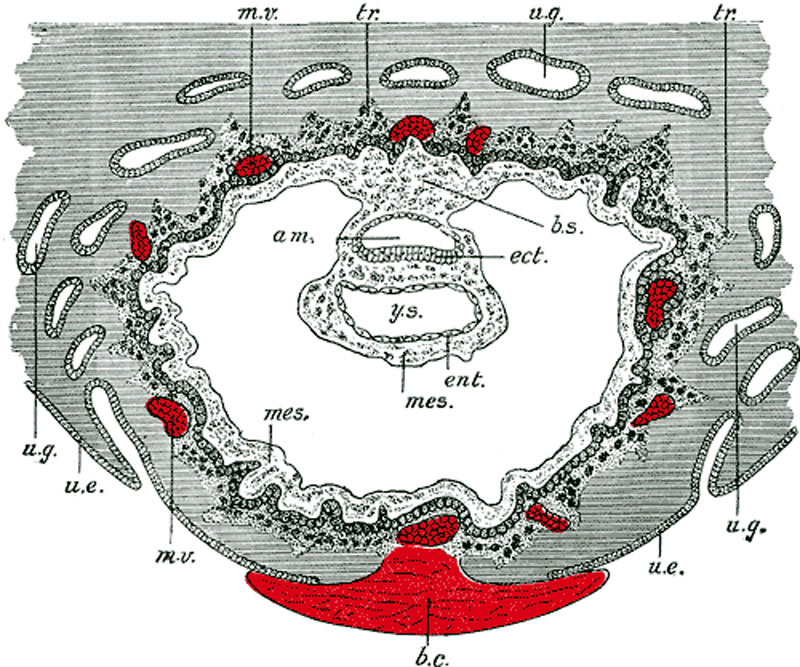
| 11:12, 22 May 2011 | Gray0032.jpg (file) |  |
159 KB | ==Human Embryo Day 8 to 9== Section through ovum imbedded in the uterine decidua. Semidiagrammatic. (After Peters.) original figure title * '''am.''' - amniotic cavity * '''b.c.''' - blood clot, at the site of initial implantation * '''b.s.''' - body-st | 1 |
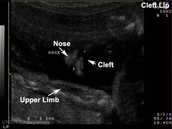
| 01:05, 18 May 2011 | Cleft lip 03.jpg (file) |  |
38 KB | 2 | |
| 00:25, 18 May 2011 | Cleft lip 02.jpg (file) |  |
22 KB | 1 | |
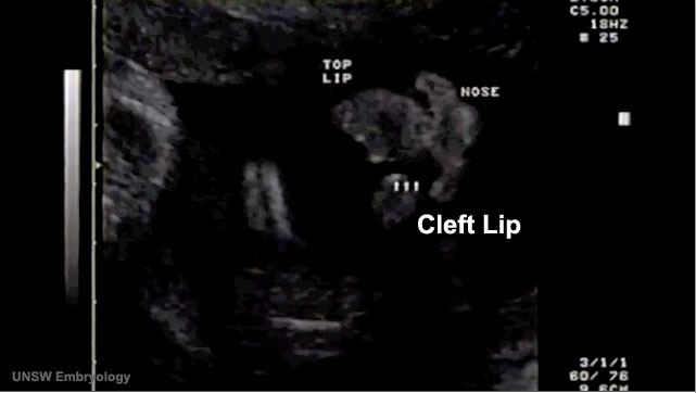
| 00:24, 18 May 2011 | Cleft lip 100.jpg (file) |  |
33 KB | ==Cleft Lip== A common form of abnormal head development is associated with clefting of the lip which may also include cleft palate. The movie above shows a fetus (at 18 weeks gestation, 20 weeks obstetric) which has a facial cleft. Measure cleft - imag | 1 |
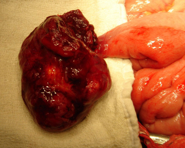
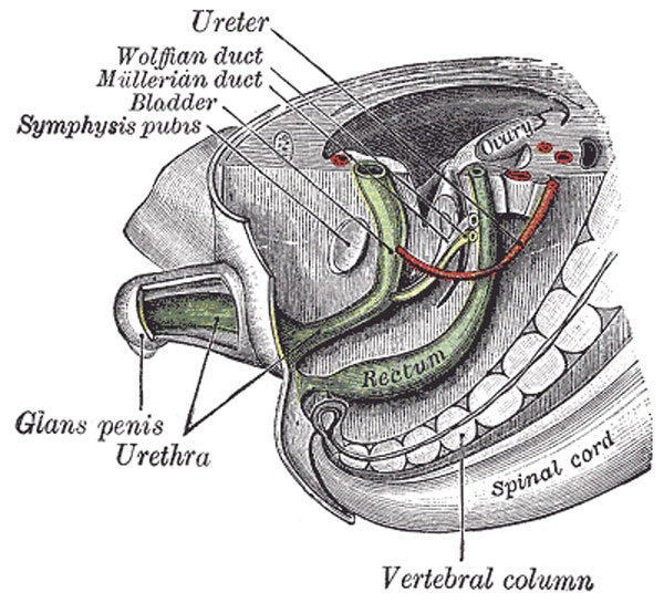
| 18:42, 29 April 2011 | Meckel's diverticulum 03.jpg (file) |  |
69 KB | Intraoperative picture showing tumour arising from Meckel's diverticulum; Tumour was located in Meckel's diverticulum and adherent to wall of urinary bladder. Meckel's Diverticulum is the most commonly encountered congenital anomaly of the small intestin | 1 |
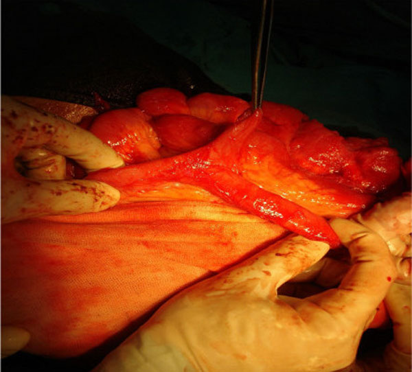
| 18:22, 29 April 2011 | Meckel's diverticulum 02.jpg (file) |  |
58 KB | ==Meckel's Diverticulum== Meckel's diverticulum is the remain of the prenatal yolkstalk (Vitellointestinal duct). The yolk sac of the developing embryo is connected to the primitive gut by the yolk stalk or vitelline (i.e. omphalomesenteric) duct. This s | 1 |
| 16:01, 28 April 2011 | Gray0993.jpg (file) |  |
88 KB | ==Week 8 Human Embryo== Tail end of human embryo, from eight and a half to nine weeks old (From model by Keibel.) The entodermal cloaca is divided into a dorsal and a ventral part by means of a partition, the urorectal septum (Fig. 992), which grows | 1 |
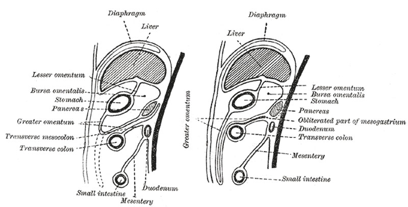
| 15:56, 28 April 2011 | Gray0990.jpg (file) |  |
60 KB | ==Diagrams to illustrate the development of the greater omentum and transverse mesocolon== Further changes take place in the bursa omentalis and in the common mesentery, and give rise to the peritoneal relations seen in the adult. The bursa omentalis, wh | 1 |
| 13:12, 15 April 2011 | Adult gastrointestinal tract cartoon02.jpg (file) |  |
42 KB | 1 | |
| 13:12, 15 April 2011 | Adult gastrointestinal tract cartoon01.jpg (file) |  |
55 KB | 1 | |
| 17:11, 5 April 2011 | Logo.png (file) |  |
21 KB | This is the image used for the website icon. Based upon Carnegie stage 23 Embryo with text superimposed in UNSW font. | 1 |
| 13:56, 14 February 2011 | Brown010.jpg (file) |  |
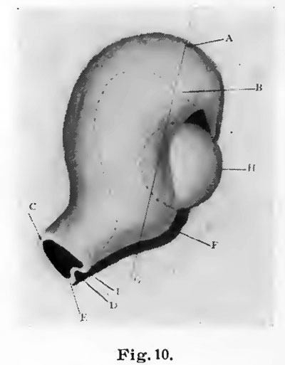
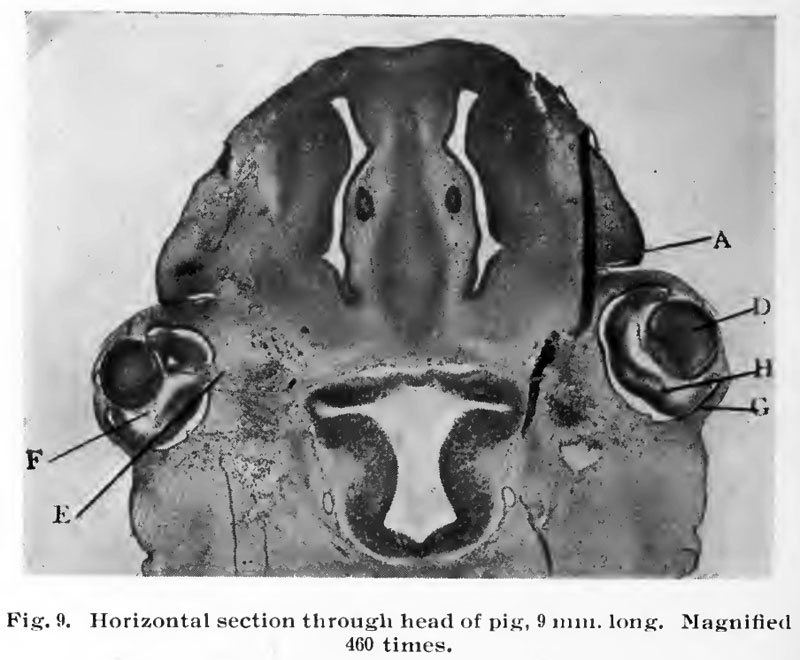
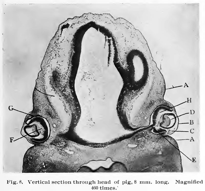
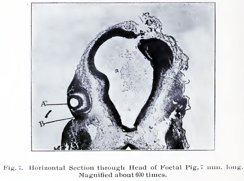
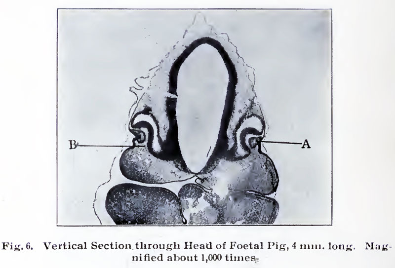
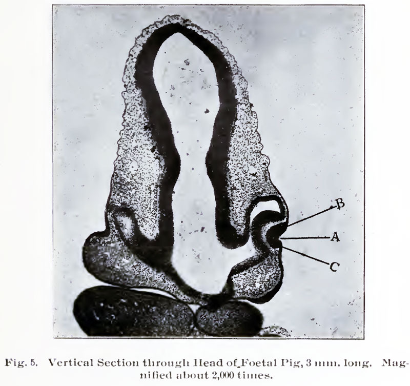
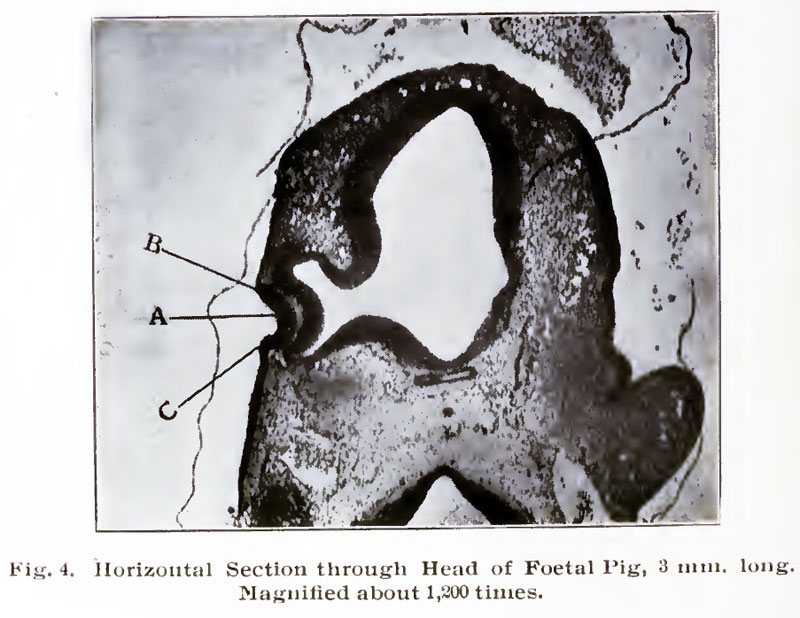
21 KB | {{Template:Brown 1906 Figures}} Category:Pig | 1 |
| 13:56, 14 February 2011 | Brown009.jpg (file) |  |
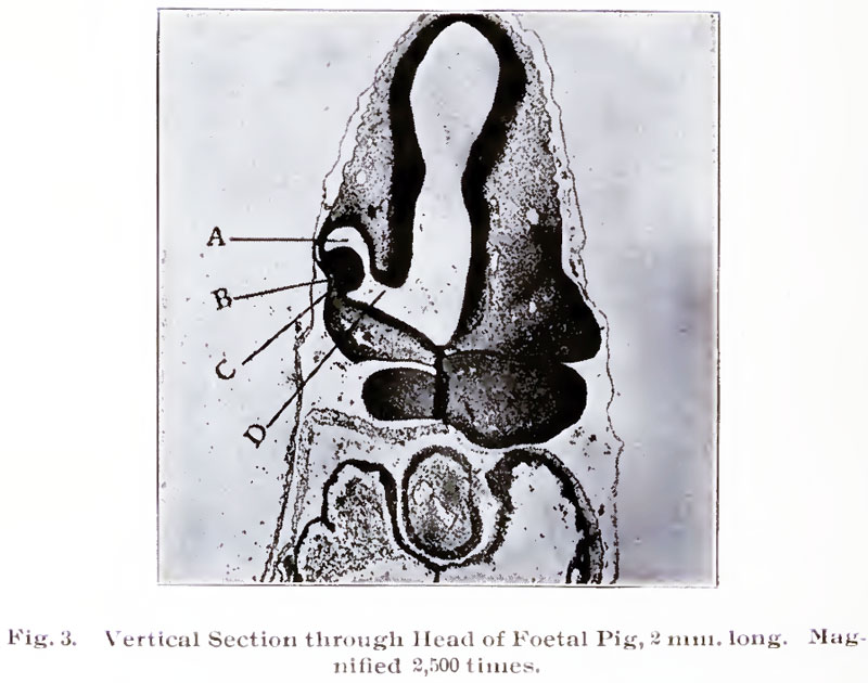
78 KB | {{Template:Brown 1906 Figures}} Category:Pig | 1 |
| 13:56, 14 February 2011 | Brown008.jpg (file) |  |
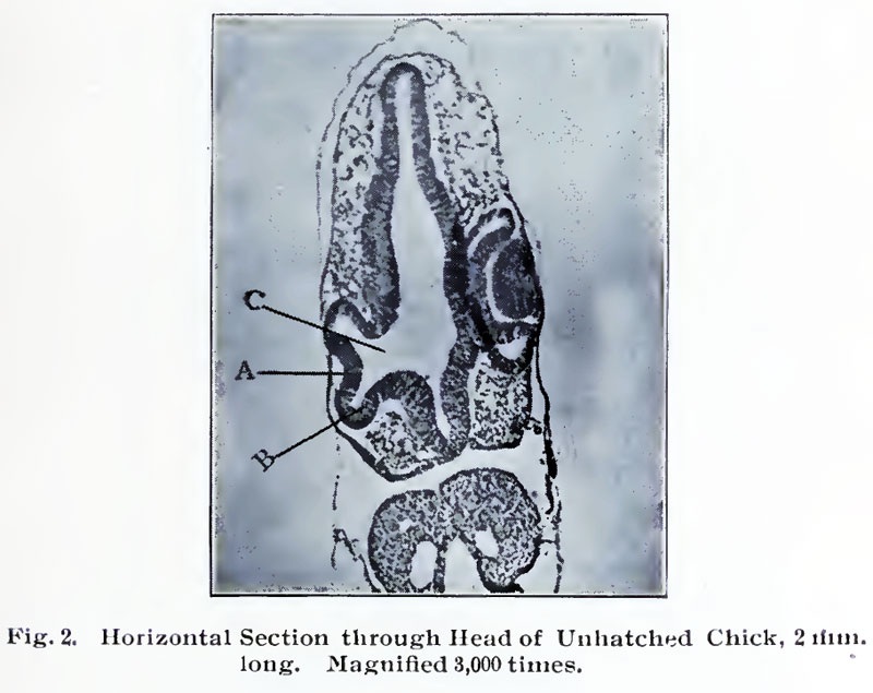
112 KB | {{Template:Brown 1906 Figures}} Category:Pig | 1 |
| 13:13, 14 February 2011 | Brown007.jpg (file) |  |
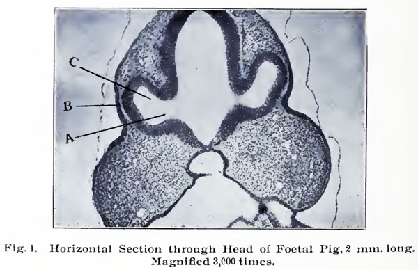
78 KB | {{Template:Brown 1906 Figures}} | 1 |
| 13:12, 14 February 2011 | Brown006.jpg (file) |  |
63 KB | {{Template:Brown 1906 Figures}} | 1 |
| 13:12, 14 February 2011 | Brown005.jpg (file) |  |
99 KB | {{Template:Brown 1906 Figures}} | 1 |
| 13:12, 14 February 2011 | Brown004.jpg (file) |  |
93 KB | {{Template:Brown 1906 Figures}} | 1 |
| 13:12, 14 February 2011 | Brown003.jpg (file) |  |
85 KB | {{Template:Brown 1906 Figures}} | 1 |
| 13:11, 14 February 2011 | Brown002.jpg (file) |  |
73 KB | {{Template:Brown 1906 Figures}} | 1 |
| 13:11, 14 February 2011 | Brown001.jpg (file) |  |
52 KB | {{Template:Brown 1906 Figures}} | 1 |
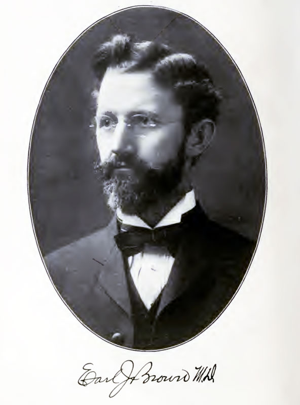
| 11:36, 14 February 2011 | Earl J. Brown.jpg (file) |  |
48 KB | ==Earl J. Brown== M. D. Professor of Histology of the Eye, at the Chicago Eye, Ear, Nose and Throat College Historic Textbook The Embryology Anatomy and Histology of the Eye | 1 |
| 08:33, 19 January 2011 | Bailey231.jpg (file) |  |
69 KB | {{Template:Bailey 1921 Figures}} Category:Human Category:Musculoskeletal | 1 |
| 08:33, 19 January 2011 | Bailey230.jpg (file) |  |
145 KB | {{Template:Bailey 1921 Figures}} Category:Human Category:Musculoskeletal | 1 |
| 08:33, 19 January 2011 | Bailey229.jpg (file) |  |
86 KB | {{Template:Bailey 1921 Figures}} Category:Human Category:Musculoskeletal | 1 |
| 08:32, 19 January 2011 | Bailey228.jpg (file) |  |
134 KB | {{Template:Bailey 1921 Figures}} Category:Human Category:Musculoskeletal | 1 |
| 08:32, 19 January 2011 | Bailey227.jpg (file) |  |
226 KB | {{Template:Bailey 1921 Figures}} Category:Human Category:Musculoskeletal | 1 |
| 08:31, 19 January 2011 | Bailey226.jpg (file) |  |
71 KB | {{Template:Bailey 1921 Figures}} Category:Human Category:Musculoskeletal | 1 |
| 08:31, 19 January 2011 | Bailey225.jpg (file) |  |
100 KB | {{Template:Bailey 1921 Figures}} Category:Human Category:Musculoskeletal | 1 |
| 08:31, 19 January 2011 | Bailey224.jpg (file) |  |
114 KB | {{Template:Bailey 1921 Figures}} Category:Human Category:Musculoskeletal | 1 |
| 08:30, 19 January 2011 | Bailey223.jpg (file) |  |
108 KB | {{Template:Bailey 1921 Figures}} Category:Human Category:Musculoskeletal | 1 |
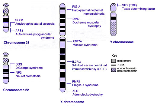
| 13:44, 25 November 2010 | Human genetics chromosomes 21-XY.jpg (file) |  |
41 KB | ==Human Genetic Disorders Chromosomes 21 to X and Y== Human genetics chromosomes 21-XY.jpg Category:Genetics Category:Genetic Abnormalities Category:Molecular | 1 |
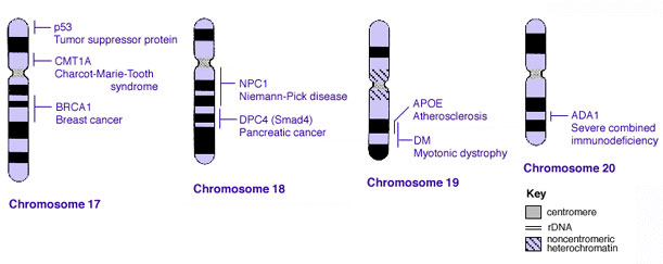
| 13:43, 25 November 2010 | Human genetics chromosomes 17-20.jpg (file) |  |
26 KB | ==Human Genetic Disorders Chromosomes 17 to 20== Human genetics chromosomes 17-20.jpg Category:Genetics Category:Genetic Abnormalities Category:Molecular | 1 |
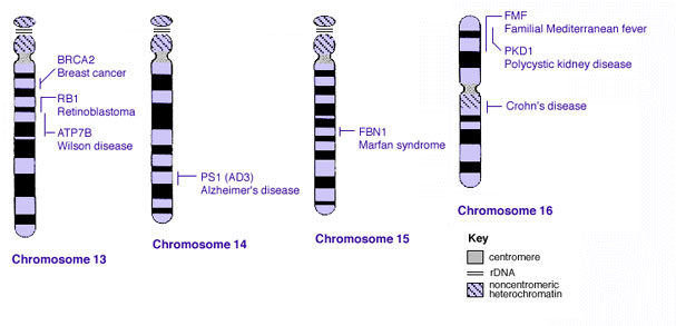
| 13:42, 25 November 2010 | Human genetics chromosomes 13-16.jpg (file) |  |
29 KB | ==Human Genetic Disorders Chromosomes 13 to 16== Human genetics chromosomes 13-16.jpg Category:Genetics Category:Genetic Abnormalities Category:Molecular | 1 |
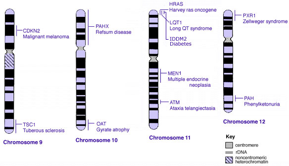
| 13:42, 25 November 2010 | Human genetics chromosomes 9-12.jpg (file) |  |
35 KB | ==Human Genetic Disorders Chromosomes 9 to 12== Human genetics chromosomes 9-12.jpg Category:Genetics Category:Genetic Abnormalities Category:Molecular | 1 |
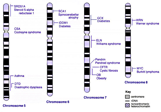
| 13:41, 25 November 2010 | Human genetics chromosomes 5-8.jpg (file) |  |
44 KB | ==Human Genetic Disorders Chromosomes 5 to 8== Human genetics chromosomes 5-8.jpg Category:Genetics Category:Genetic Abnormalities Category:Molecular | 1 |
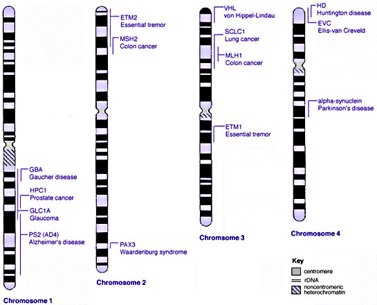
| 13:39, 25 November 2010 | Human genetics chromosomes 1-4.jpg (file) |  |
44 KB | ==Human Genetic Disorders Chromosomes 1 to 4== Human genetics chromosomes 1-4.jpg Category:Genetics Category:Genetic Abnormalities Category:Molecular | 1 |
| 11:45, 8 May 2010 | Podcast icon.jpg (file) | 2 KB | Podcast icon | 1 | |
| 18:13, 14 April 2010 | Sow and piglet.jpg (file) |  |
43 KB | 2 | |
| 18:28, 19 October 2009 | Stage16-18 face 01 icon.jpg (file) | 4 KB | 1 | ||
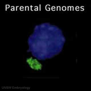
| 18:11, 19 October 2009 | Parental genome mix 01 icon.jpg (file) | 4 KB | 1 | ||
| 17:52, 19 October 2009 | Blastomere mitosis 01 icon.jpg (file) | 3 KB | 1 | ||
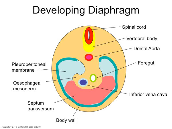
| 15:49, 24 August 2009 | Diaphragm components.jpg (file) |  |
41 KB | Diaphragm components cartoon Image Source: UNSW Embryology http://embryology.med.unsw.edu.au/Notes/images/humanlung/diaphragm1.jpg | 1 |
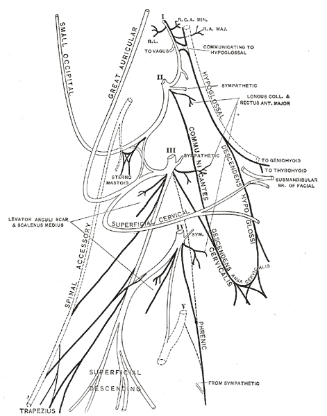
| 15:48, 24 August 2009 | Gray804.gif (file) |  |
30 KB | Adult Cervical Plexus (phrenic nerve shown lower right) nnervation of the human diaphragm is by the phrenic nerves, arising from the same segmental levels from which the diaphragm skeletal muscles arise, segmental levels C3 to C5. The paired phrenic n | 1 |
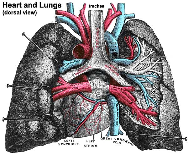
| 14:35, 24 August 2009 | Historic-lungs.jpg (file) |  |
79 KB | Historic drawing of the lungs showing dorsal view and anatomical size and position with respect to the heart. Category:Respiratory Category:Historic Category:Gray's 1918 Anatomy | 1 |
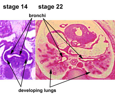
| 14:28, 24 August 2009 | Stage14-22 lungs.jpg (file) |  |
55 KB | 1 | |
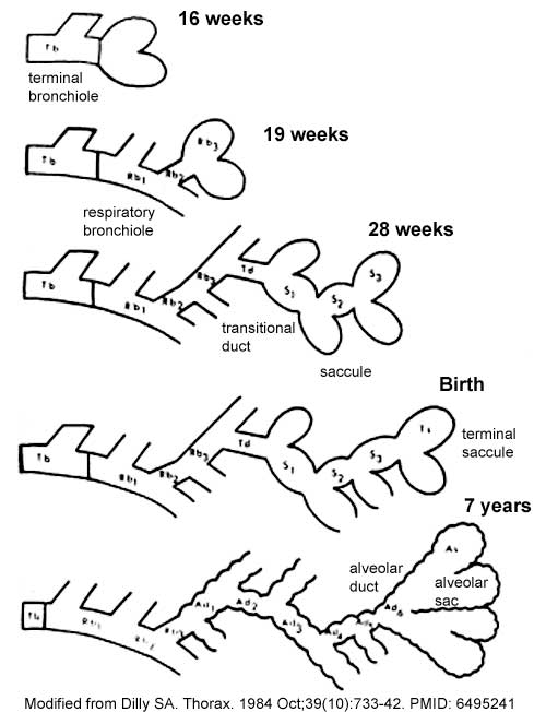
| 14:10, 24 August 2009 | Lung alveoli development cartoon.jpg (file) |  |
38 KB | Lung alveoli development Image modified from: Scanning electron microscope study of the development of the human respiratory acinus. Dilly SA. Thorax. 1984 Oct;39(10):733-42. [http://www.ncbi.nlm.nih.gov/pubmed/6495241 PMID: 6495241] Thorax 1984;39:733 | 1 |