Uploads by MarkHill
From Embryology
This special page shows all uploaded files.
| Date | Name | Thumbnail | Size | Description | Versions |
|---|---|---|---|---|---|
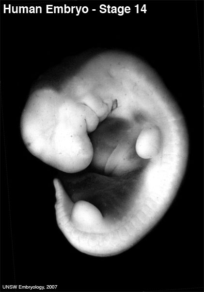
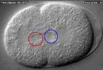
| 20:49, 13 May 2009 | 600px stage14.jpg (file) |  |
27 KB | Human Embryo stage 14 Image source: UNSW Embryology © Dr Mark Hill, Image cannot be reproduced without permission. | 1 |
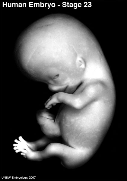
| 20:49, 13 May 2009 | 600px stage23.jpg (file) |  |
26 KB | Human Embryo stage 23 Image source: UNSW Embryology © Dr Mark Hill, Image cannot be reproduced without permission. | 1 |
| 11:42, 12 August 2009 | Abnormal81-92-neuron.png (file) |  |
9 KB | Pie diagram shows the percentage of neural defects of all notifiable birth defects in Australia. Data groupings and classification as Major or Minor Abnormalities are based on that used by the Australian Institute of Health and Welfare National Perinatal | 1 |
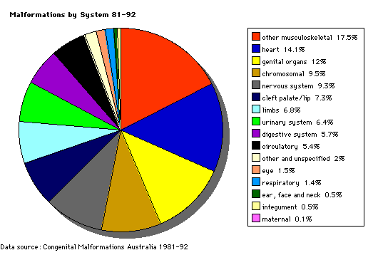
| 14:13, 12 August 2009 | Abnormal AusData81-92.png (file) |  |
10 KB | Pie diagram shows the percentage of developmental abnormalities by categories of all notifiable birth defects in Australia. Data groupings and classification as Major or Minor Abnormalities are based on that used by the Australian Institute of Health and | 1 |
| 14:16, 12 August 2009 | Abnormal AusData81-92Graph.png (file) |  |
7 KB | Pie diagram shows the percentage of developmental abnormalities by categories of all notifiable birth defects in Australia. Data groupings and classification as Major or Minor Abnormalities are based on that used by the Australian Institute of Health and | 1 |
| 13:12, 15 April 2011 | Adult gastrointestinal tract cartoon01.jpg (file) |  |
55 KB | 1 | |
| 13:12, 15 April 2011 | Adult gastrointestinal tract cartoon02.jpg (file) |  |
42 KB | 1 | |
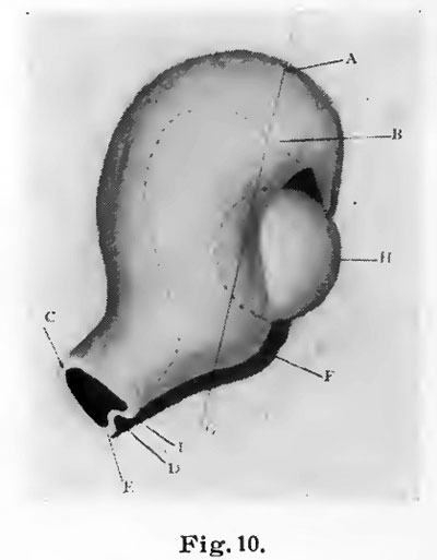
| 08:30, 19 January 2011 | Bailey223.jpg (file) |  |
108 KB | {{Template:Bailey 1921 Figures}} Category:Human Category:Musculoskeletal | 1 |
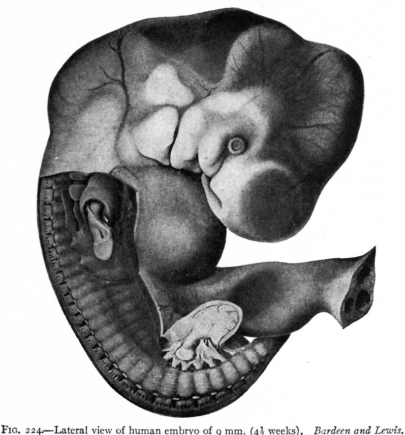
| 08:31, 19 January 2011 | Bailey224.jpg (file) |  |
114 KB | {{Template:Bailey 1921 Figures}} Category:Human Category:Musculoskeletal | 1 |
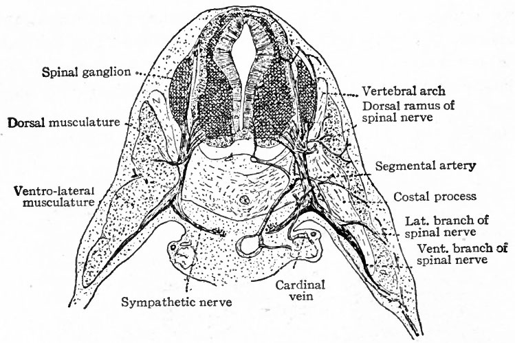
| 08:31, 19 January 2011 | Bailey225.jpg (file) |  |
100 KB | {{Template:Bailey 1921 Figures}} Category:Human Category:Musculoskeletal | 1 |
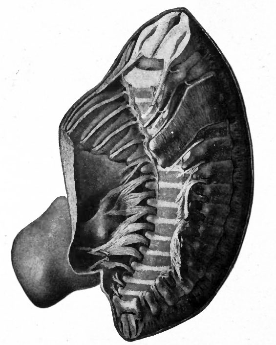
| 08:31, 19 January 2011 | Bailey226.jpg (file) |  |
71 KB | {{Template:Bailey 1921 Figures}} Category:Human Category:Musculoskeletal | 1 |
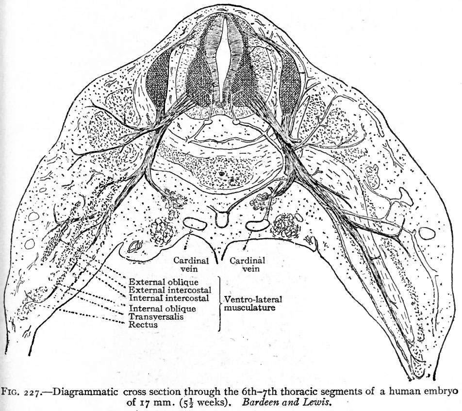
| 08:32, 19 January 2011 | Bailey227.jpg (file) |  |
226 KB | {{Template:Bailey 1921 Figures}} Category:Human Category:Musculoskeletal | 1 |
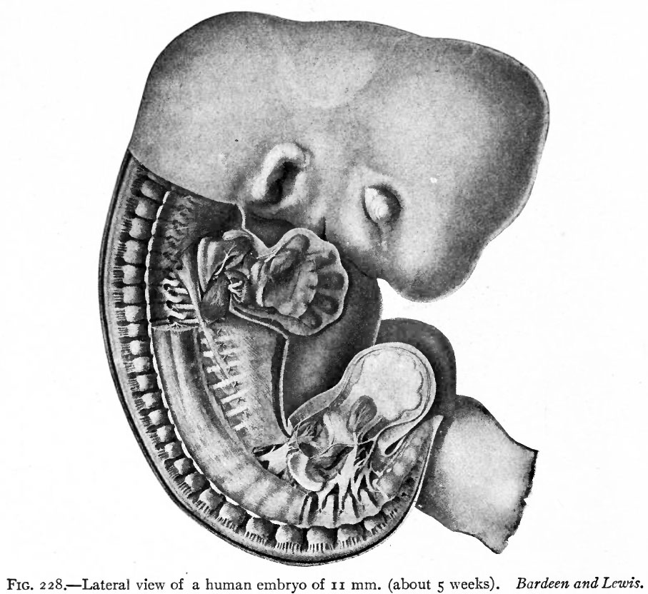
| 08:32, 19 January 2011 | Bailey228.jpg (file) |  |
134 KB | {{Template:Bailey 1921 Figures}} Category:Human Category:Musculoskeletal | 1 |
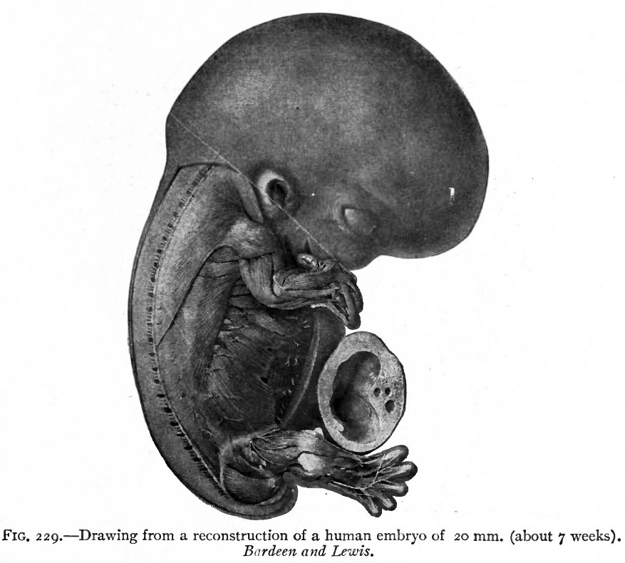
| 08:33, 19 January 2011 | Bailey229.jpg (file) |  |
86 KB | {{Template:Bailey 1921 Figures}} Category:Human Category:Musculoskeletal | 1 |
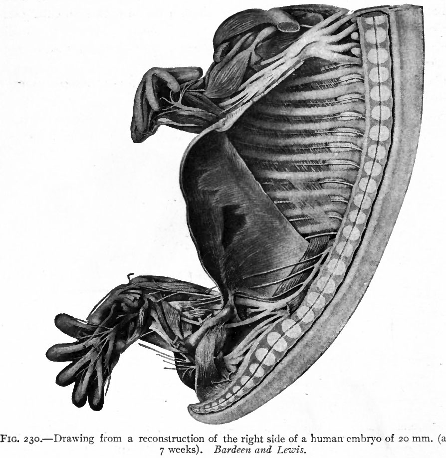
| 08:33, 19 January 2011 | Bailey230.jpg (file) |  |
145 KB | {{Template:Bailey 1921 Figures}} Category:Human Category:Musculoskeletal | 1 |
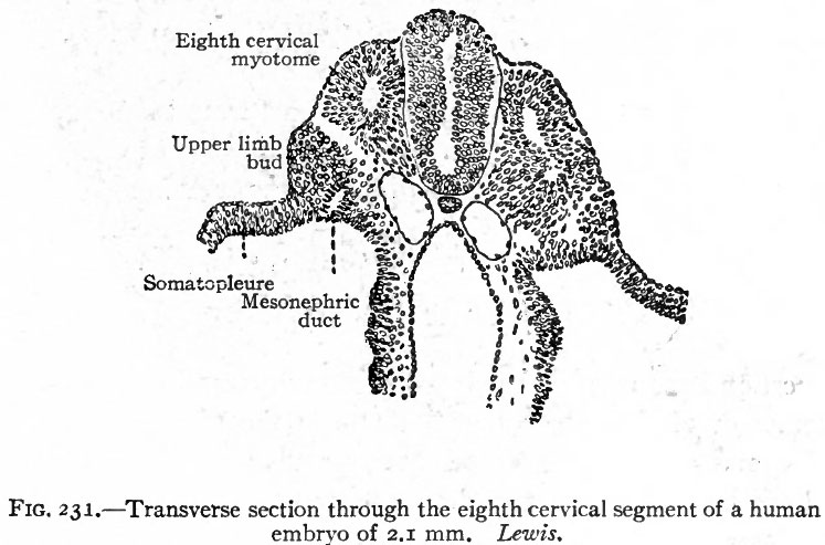
| 08:33, 19 January 2011 | Bailey231.jpg (file) |  |
69 KB | {{Template:Bailey 1921 Figures}} Category:Human Category:Musculoskeletal | 1 |
| 12:20, 20 August 2009 | Beverley hill.jpg (file) |  |
3 KB | Beverley Hill (1933 - 2001) who from 1996 - 2000 worked patiently on all facets of this project; preparing images, fixing text, links, and committing a tireless labour on my continual updates and changes. | 1 |
| 17:52, 19 October 2009 | Blastomere mitosis 01 icon.jpg (file) | 3 KB | 1 | ||
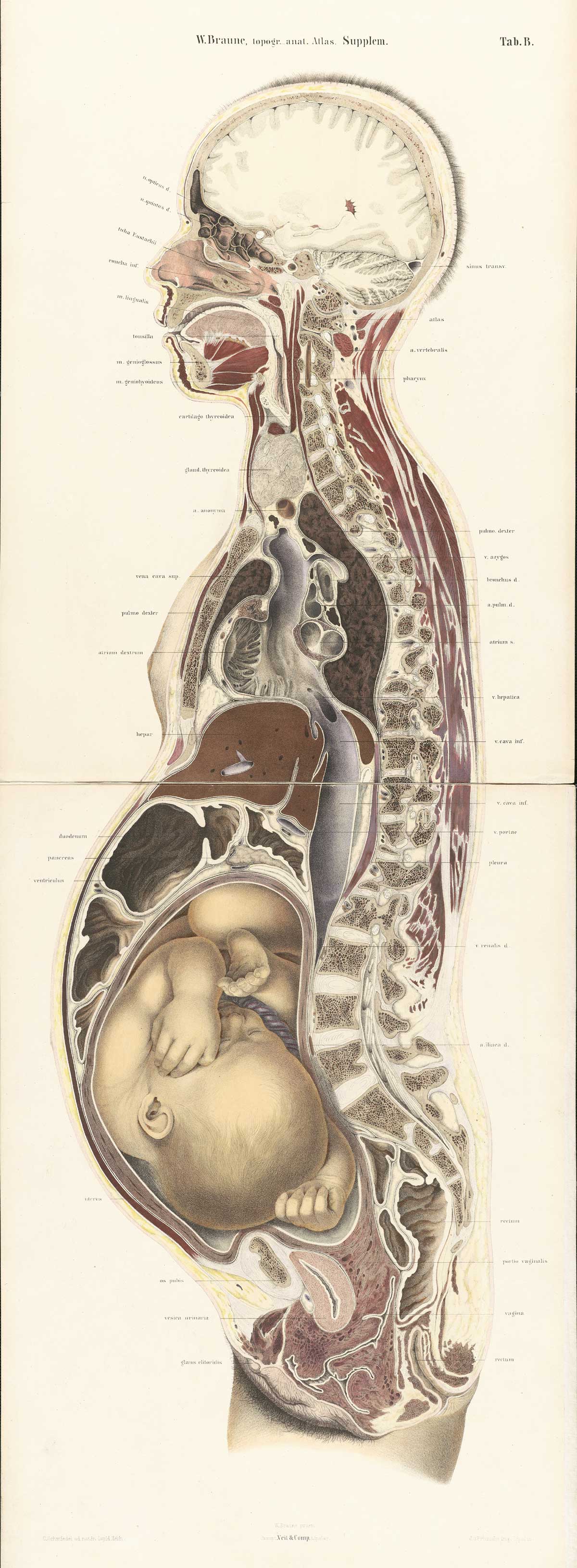
| 15:18, 22 July 2009 | Braune2 B1.jpg (file) |  |
413 KB | 1 | |
| 13:11, 14 February 2011 | Brown001.jpg (file) |  |
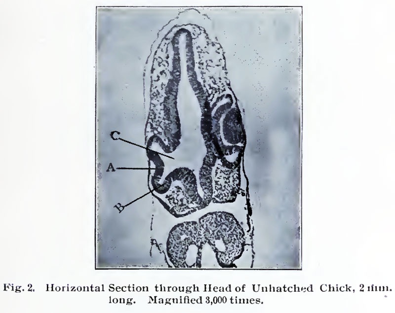
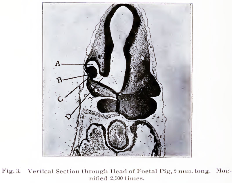
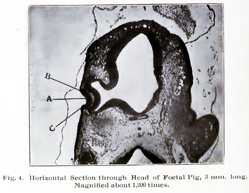
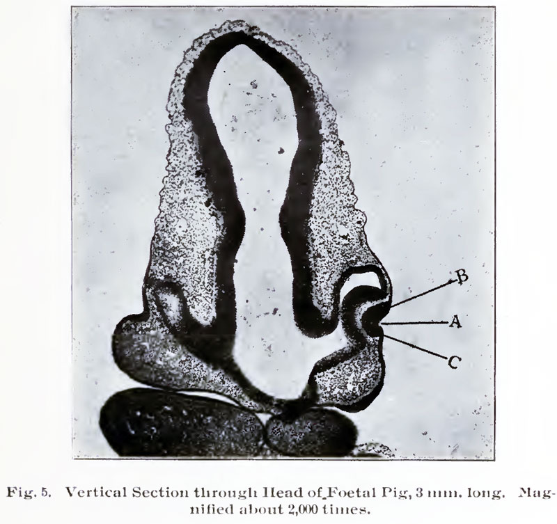
52 KB | {{Template:Brown 1906 Figures}} | 1 |
| 13:11, 14 February 2011 | Brown002.jpg (file) |  |
73 KB | {{Template:Brown 1906 Figures}} | 1 |
| 13:12, 14 February 2011 | Brown003.jpg (file) |  |
85 KB | {{Template:Brown 1906 Figures}} | 1 |
| 13:12, 14 February 2011 | Brown004.jpg (file) |  |
93 KB | {{Template:Brown 1906 Figures}} | 1 |
| 13:12, 14 February 2011 | Brown005.jpg (file) |  |
99 KB | {{Template:Brown 1906 Figures}} | 1 |
| 13:12, 14 February 2011 | Brown006.jpg (file) |  |
63 KB | {{Template:Brown 1906 Figures}} | 1 |
| 13:13, 14 February 2011 | Brown007.jpg (file) |  |
78 KB | {{Template:Brown 1906 Figures}} | 1 |
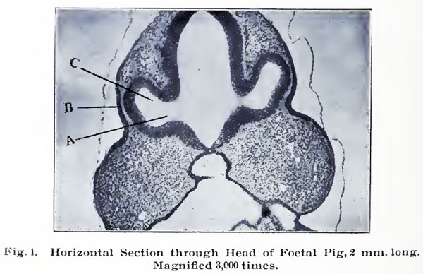
| 13:56, 14 February 2011 | Brown008.jpg (file) |  |
112 KB | {{Template:Brown 1906 Figures}} Category:Pig | 1 |
| 13:56, 14 February 2011 | Brown009.jpg (file) |  |
78 KB | {{Template:Brown 1906 Figures}} Category:Pig | 1 |
| 13:56, 14 February 2011 | Brown010.jpg (file) |  |
21 KB | {{Template:Brown 1906 Figures}} Category:Pig | 1 |
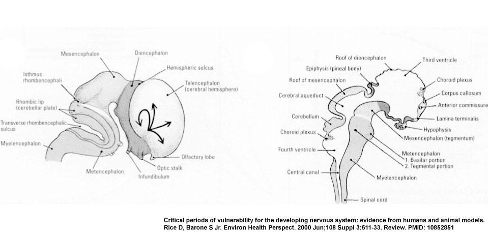
| 01:07, 11 August 2009 | CNS later development.jpg (file) |  |
59 KB | CNS later development cartoon (E) The lateral view shows the migratory paths from the more central ventricular zone and gradients maturation of the neocortex (see arrows). (F) The midsagittal view of the brain and spinal cord, with the major divisions d | 1 |
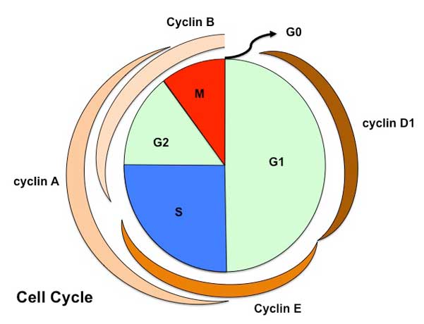
| 15:16, 27 July 2009 | Cell cycle1.jpg (file) |  |
23 KB | Cartoon of Cell Cycle (large) Showing phases and cyclin levels Source: Mark Hill | 1 |
| 15:12, 20 January 2009 | Chick15h.jpg (file) |  |
3 KB | chicken embryo 15 hours from fertilization. | 1 |
| 15:54, 21 July 2009 | Chris.gif (file) |  |
13 KB | Large embryology icon Image | 1 |
| 12:08, 20 August 2009 | Chris.jpg (file) |  |
6 KB | UNSW Embryology logo | 1 |
| 15:36, 6 August 2009 | Citeulike 16x16.png (file) |  |
413 bytes | 1 | |
| 00:25, 18 May 2011 | Cleft lip 02.jpg (file) |  |
22 KB | 1 | |
| 01:05, 18 May 2011 | Cleft lip 03.jpg (file) |  |
38 KB | 2 | |
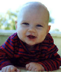
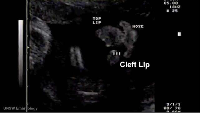
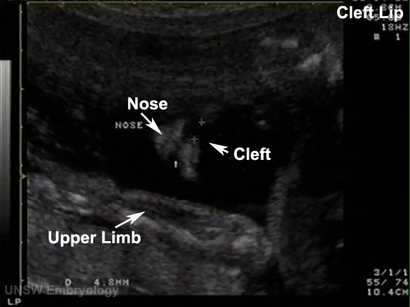
| 00:24, 18 May 2011 | Cleft lip 100.jpg (file) |  |
33 KB | ==Cleft Lip== A common form of abnormal head development is associated with clefting of the lip which may also include cleft palate. The movie above shows a fetus (at 18 weeks gestation, 20 weeks obstetric) which has a facial cleft. Measure cleft - imag | 1 |
| 15:37, 6 August 2009 | Connotea 32x32.png (file) |  |
1 KB | 1 | |
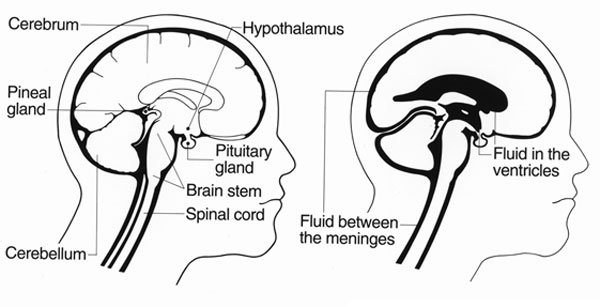
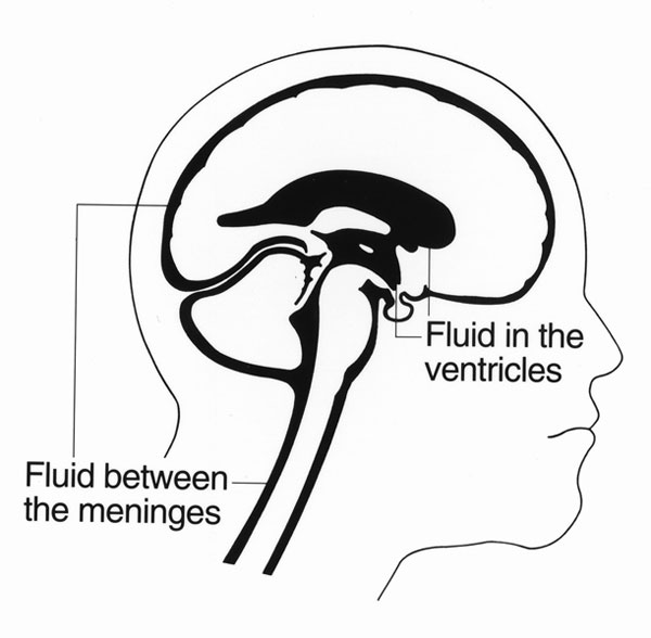
| 15:06, 10 August 2009 | Csf cartoon1.jpg (file) |  |
35 KB | Cerebrospinal fluid (CSF) location in the brain and spinal cord (CNS) - cartoon | 1 |
| 15:07, 10 August 2009 | Csf cartoon2.jpg (file) |  |
44 KB | Cerebrospinal fluid (CSF) location in the brain and spinal cord (CNS) - cartoon | 1 |
| 15:07, 10 August 2009 | Csf cartoon3.jpg (file) |  |
37 KB | 1 | |
| 15:35, 6 August 2009 | Delicious 32x32.png (file) |  |
888 bytes | 1 | |
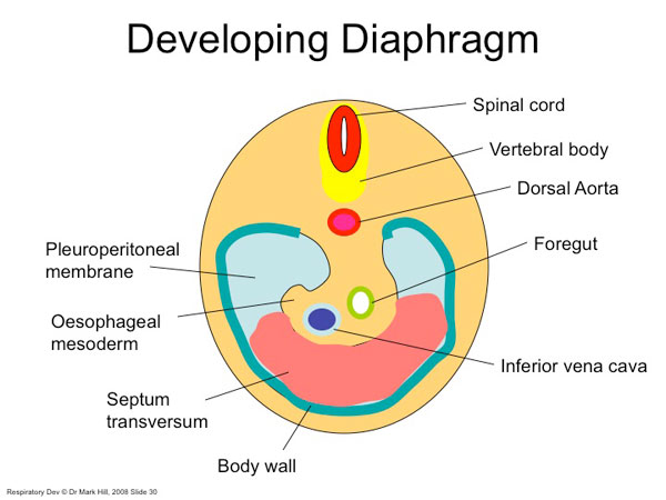
| 15:49, 24 August 2009 | Diaphragm components.jpg (file) |  |
41 KB | Diaphragm components cartoon Image Source: UNSW Embryology http://embryology.med.unsw.edu.au/Notes/images/humanlung/diaphragm1.jpg | 1 |
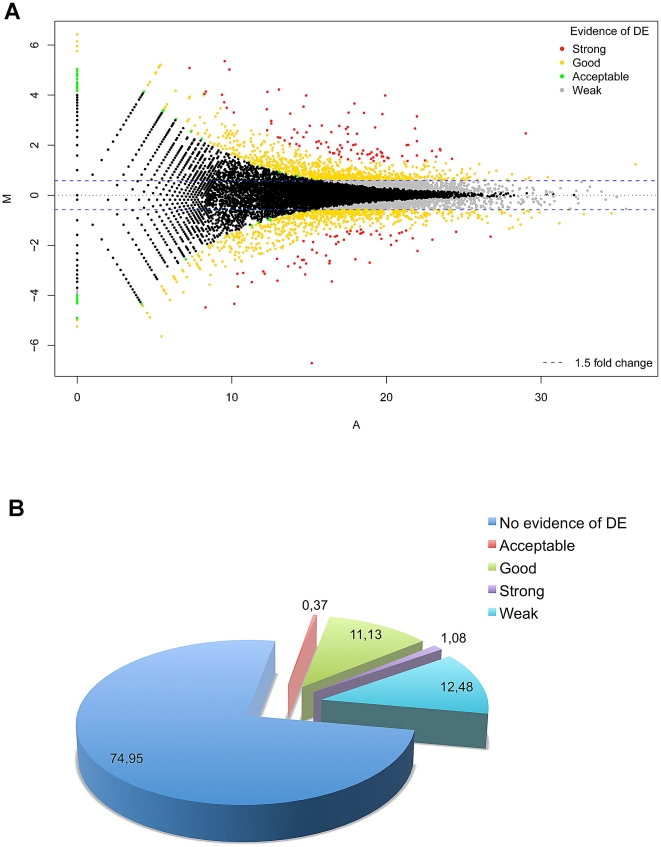
| 18:33, 11 August 2011 | Differentially expressed RefSeq genes in human trisomy 21.jpg (file) |  |
171 KB | Reverted to version as of 02:46, 11 August 2011 | 3 |
| 15:34, 6 August 2009 | Digg 32x32.png (file) |  |
2 KB | 1 | |
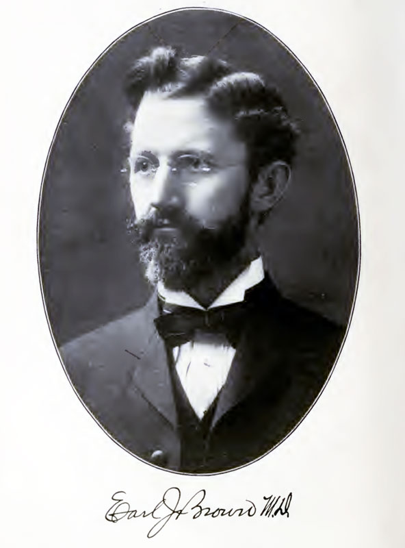
| 11:36, 14 February 2011 | Earl J. Brown.jpg (file) |  |
48 KB | ==Earl J. Brown== M. D. Professor of Histology of the Eye, at the Chicago Eye, Ear, Nose and Throat College Historic Textbook The Embryology Anatomy and Histology of the Eye | 1 |
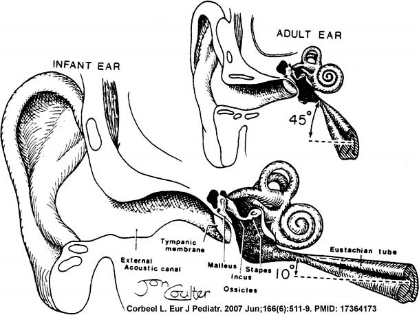
| 23:06, 13 May 2009 | Eustacian tube angle.jpg (file) |  |
48 KB | Newborn to adult Eustachian (auditory, otopharyngeal or pharyngotympanic) tube. Connects middle ear cavity to nasopharynx portion of pharynx Ventilation - pressure equalization in the middle ear Clearance - allow fluid drainage from the middle ear Tube | 1 |
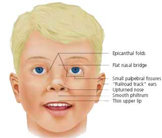
| 23:10, 13 May 2009 | FASface.jpg (file) |  |
8 KB | Facial Appearance of FAS Some, or all, of the following facial features are associated with FAS * Microcephaly - leads to small head circumference * Palpebral fissure - short opening of eye * Epicanthal folds - fold of skin at inside of corner of eye * M | 1 |
| 15:33, 6 August 2009 | Facebook 16x16.png (file) |  |
1 KB | 1 |