Foundations - Histology Cells and Tissues
Introduction


Background and Self-directed Learning for Medicine Foundations.
Practical - Histology Cells and Tissues Virtual Slides by Patrick de Permentier.
This current page content is not part of the Foundations practical class.
- Links: Histology Virtual Slides | Histology Introduction | Histology Epithelia and Skin | Histology Stains | Histology Drawings
Specific Objectives
- Obtain an understanding of the histological appearance of the basic tissues namely epithelium, connective tissue, muscle and nervous tissue.
- To examine unique cellular characteristics of each of the basic tissues.
Introduction
A tissue is a functional aggregation of similar cells and their intercellular materials that combine to perform common functions. An organ is an anatomically discrete structure (e.g. heart, skin) with 1 or more functions.
Four tissues are considered basic or primary: epithelial, connective, muscular and nervous. Many organs contain all 4 types of tissues e.g. skin (covering, packing, muscles, nerves).
Epithelium
Epithelium forms continuous layers of cells that cover surfaces and line cavities of the body. These cavities include the closed peritoneal, pleural, and pericardial cavities, where the epithelium is called mesothelium, and open organ cavities, i.e. digestive, respiratory, and urogenital organs, which connect with the outside. In addition, epithelium lines the cardiovascular and lymph passageways as endothelium.
The parenchymal (secretory) cells of glands (e.g. sweat, salivary) are also epithelium. Epithelial cells are always in close apposition to each other, with a space between membranes of only about 20nm. A small amount of intercellular material, called cement substance (glycosaminoglycan) allows cells to glide over each other and offers only minimal resistance to the migration of leukocytes and other connective tissue cells through the epithelial layers. Epithelial attachment points (junctional complexes) occur between neighboring epithelial cells to hold adjacent cell membranes in close apposition. They also serve as anchoring sites for the fine filaments of the cytoskeleton, which assists in stabilizing the cell shape.
Epithelial cells rest on a basal lamina separating them from underlying connective tissue (CT). Epithelium is avascular and for its nutrition depends on diffusion of substances across the basement membrane.
Simple Epithelium
VIRTUAL SLIDE: Gallbladder eg simple epithelium


Stratified Epithelium
VIRTUAL SLIDE: Oesophagus eg stratified epithelium
|

Oesophagus stratified epithelium |
Glands

Glands are invaginations of epithelial surfaces that are formed during embryonic development by proliferation of epithelium into the underlying connective tissues to form secretory units. The secretory units, along with their ducts, are the parenchyma of the gland; the stroma of the gland represents the elements of the connective tissue that support the parenchyma. Some glands (endocrine) loose the duct and secrete directly into the blood (e.g. hormones).
VIRTUAL SLIDE: Skin


Connective Tissue

Connective tissues (CT) are the supporting framework for all tissues and organs of the body. In addition, they provide the means of anchoring and binding organs together as well as providing the packing tissue between them. CT varies in structure and character from loose (subcutaneous areas), to dense irregular (dermis of skin), to dense regular (ligaments and tendons), to firm and flexible (cartilage in the trachea, intervertebral discs, external ear, to rigid (calcified bone), to circulating (blood and lymph). Despite this wide range, all CT have an intercellular matrix composed of an amorphous ground substance in which are embedded cells and one or more types of extracellular fibres, (elastic, collagen, or reticular). In blood and lymph, the fibres are strands of fibrin, seen only during clotting. The predominant cell type of CT is the fibroblast which makes fibres. Other types are wandering cells such as mast cells, plasma cells, various leukocytes, and macrophages.
VIRTUAL SLIDES: Lymph node-silver stain, Lymph node (H&E) and Skin.
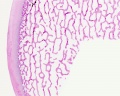
VIRTUAL SLIDE: Hyaline cartilage in trachea.
VIRTUAL SLIDE: Decalcified rib.
Bone (trabecular)
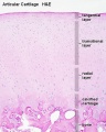
Articular cartilage


Muscle
Muscle cells (fibers; the cell is longer than wide) produce force which can be used for movements such as locomotion, contraction of organs (e.g. bladder) and pumping movements of the body. This is achieved by the muscle cells contractility state by changing their length and developing tension. The contractile elements of muscle cells are (myofibrils) composed of specific arrays of myofilaments, the proteins (actin and myosin) responsible for the contractile capability of the cell.
VIRTUAL SLIDES: Skeletal-smooth-cardiac muscle and Tongue.
Skeletal Muscle
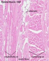
Human Skeletal Muscle (HE x4 longitudinal and transverse)
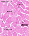
Human Skeletal Muscle (HE x40 transverse)


- Muscle Histology: Muscle Development | Human HE x4 longitudinal and transverse | Human HE x40 transverse | Human HE x40 longitudinal | Human HE x40 longitudinal | Human HE x4 longitudinal and transverse | Muscle Spindle HE x40 | Human HE x40 | Human HE x40 | Human HE x40 | Human HE x100 | Human HE x100 | Fetal human muscle | Myotendinous junction label | Myotendinous junction HE x40 | Whipf 1 | Whipf 2 | Whipf 3 | Tongue HE x10 transverse | Tongue x100 | Muscle spindle HE x20 | Muscle spindle HE x40
Smooth muscle
- Smooth muscle makes up the visceral or involuntary muscle.
- consists of spindle shaped cells of variable size.
- largest smooth muscle cells occur in the uterus during pregnancy (12x600 µm).
- smallest smooth muscle cells are found around small arterioles (1x10 µm).
- form layers surrounding the gastrointestinal tract.
- smooth muscle cells contain one centrally placed nucleus.
- innervation of smooth muscle is provided by the autonomic nervous system.
Cardiac Muscle


- Links: Heart Histology | Cardiac AZB Labeled | Cardiac AZB | Cardiac label LS | Cardiac LS | Cardiac label TS | Cardiac TS | Purkinje fibres | Purkinje fibres detail | Histology
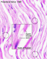
Nervous Tissue

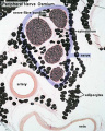
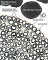
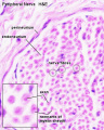
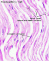
The brain and spinal cord comprise the central nervous system (CNS). The nerves that emerge from the spinal cord and brain to pass to parts of the body are the peripheral nervous tissue (PNS). Nervous tissue, with many interconnections, forms a complex system of neuronal communication within the body and is specialized for detecting stimuli, integrating functions, controlling effectors and higher functions. Nervous tissue consists of cell bodies, cell processes (nerves), and neuroglia (supporting cells).
VIRTUAL SLIDE: Peripheral Nerve.
H&E
H&E
H&E


Terms
(draft only - would need to add a brief description for each term)
- adipose - fat cells, "chicken-wire" appearance, lipid has been lost during histology processing. Two main types white adipose in many different body tissues (seen in most of your histology slides) and brown adipose for heat and energy production (seen in newborn or neonatal tissues).
- cardiac muscle - striations, intercalated discs
- collagen - the main protein found in the ECM and abundant in connective tissue. One of the most abundant proteins in the body.
- connective tissue
- epithelium
- histology
- marrow - (bone marrow)
- matrix - (extracellular matrix, ECM) material secreted by cells and lying outside cells. Connective tissue has lots of ECM between cells. Epithelia have a very little ECM, and a specialised ECM that the basal cells sit upon.
- muscle - specialised contractile cells. Three main types (smooth, skeletal and cardiac) and also characterised as striated (skeletal, cardiac) and non-striated (smooth) due to the organisation of the contractile apparatus within the muscle cell.
- periosteum - connective tissue covering the surface of bone (except articular surfaces).
- skeletal muscle - striations, fibers (muscle fiber = muscle cell), multinucleated, clustered fasicles.
- smooth muscle - no striations, sheets, elongated nuclei, involuntary (you don't control it).
Glossary Links
- Glossary: A | B | C | D | E | F | G | H | I | J | K | L | M | N | O | P | Q | R | S | T | U | V | W | X | Y | Z | Numbers | Symbols | Term Link
Cite this page: Hill, M.A. (2024, May 18) Embryology Foundations - Histology Cells and Tissues. Retrieved from https://embryology.med.unsw.edu.au/embryology/index.php/Foundations_-_Histology_Cells_and_Tissues
- © Dr Mark Hill 2024, UNSW Embryology ISBN: 978 0 7334 2609 4 - UNSW CRICOS Provider Code No. 00098G