File:X-ray Abdomen bilateral nephrocalcinosis.jpg
Original file (490 × 649 pixels, file size: 72 KB, MIME type: image/jpeg)
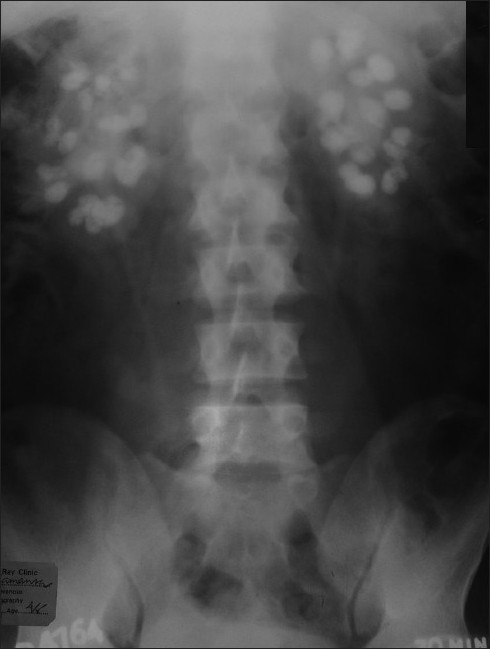
Abdominal X-ray showing extensive bilateral nephrocalcinosis
Original File Name: ASM-30-497-g004.jpg
Original Image Legend: Figure 4 Photograph shows straight X-ray of abdomen with bilateral extensive nephrocalcinosis.
Reference
<pubmed>21060166</pubmed>
PMID: 21060166
http://www.saudiannals.net/text.asp?2010/30/6/497/72282
This is an open-access article distributed under the terms of the Creative Commons Attribution License, which permits unrestricted use, distribution, and reproduction in any medium, provided the original work is properly cited.
Assessment
+ Clinically relevant image. - No explanation or labelling of "nephrocalcinosis". Project is intended for university student and should have an explanation of this clinical term and what is shown in the X-ray.
- Note - This image was originally uploaded as part of an undergraduate science student project and may contain inaccuracies in either description or acknowledgements. Students have been advised in writing concerning the reuse of content and may accidentally have misunderstood the original terms of use. If image reuse on this non-commercial educational site infringes your existing copyright, please contact the site editor for immediate removal.
File history
Click on a date/time to view the file as it appeared at that time.
| Date/Time | Thumbnail | Dimensions | User | Comment | |
|---|---|---|---|---|---|
| current | 23:32, 10 October 2011 | | 490 × 649 (72 KB) | Z3331469 (talk | contribs) | Abdominal X-ray showing extensive bilateral nephrocalcinosis Nandini Chakrabarti and Chandan Chattopadhyay [Dysparathyroidism: A Clinical Window] Ann Saudi Med. 2010 Nov-Dec; 30(6): 497–498. PMID: 21060166 Original File Name: ASM-30-497-g004.jpg Orig |
You cannot overwrite this file.
File usage
The following 2 pages use this file: