File:Uterine and placental vasculature.jpg: Difference between revisions
| Line 8: | Line 8: | ||
:'''Links:''' [[:File:Placenta_spiral_artery_conversion.jpg|Figure - Placenta spiral artery conversion]] | :'''Links:''' [[:File:Uterine_and_placental_vasculature.jpg|Figure - Uterine and placental vasculature]] | [[:File:Placenta_spiral_artery_conversion.jpg|Figure - Placenta spiral artery conversion]] | [[Placenta Development]] | ||
Original File name: Gr2.jpg | Original File name: Gr2.jpg | ||
Revision as of 15:41, 14 August 2011
Uterine and Placental Vasculature in Non-pregnant, Pregnant and immediate Post-partum State
Diagrammatic representation of uterine and placental vasculature (red shading = arterial; blue shading = venous) in the non-pregnant, pregnant and immediate post-partum state.
Normal pregnancy is characterized by the formation of large arterio-venous shunts that persist in the immediate post-partum period.
By contrast pregnancies complicated by severe preeclampsia are characterized by minimal arterio-venous shunts, and thus narrower uterine arteries. Extravillous cytotrophoblast invasion in normal pregnancy (diamonds) extends beyond the decidua into the inner myometrium resulting in the formation of funnels at the discharging tips of the spiral arteries. Contrast with severe preeclampsia. (Prepared by Ms. Leslie Proctor, MSc.)
- Links: Figure - Uterine and placental vasculature | Figure - Placenta spiral artery conversion | Placenta Development
Original File name: Gr2.jpg
Reference
<pubmed>19375795</pubmed>| PMC2697319
Placenta. 2009 June; 30(6): 473–482. doi: 10.1016/j.placenta.2009.02.009.
Copyright © 2009 Elsevier Ltd. “This is an unofficial translation of an article that appeared in an Elsevier publication. Elsevier has not endorsed this translation.”
http://www.elsevier.com/wps/find/authorsview.authors/supplementalterms1.0
File history
Click on a date/time to view the file as it appeared at that time.
| Date/Time | Thumbnail | Dimensions | User | Comment | |
|---|---|---|---|---|---|
| current | 09:43, 16 August 2009 | 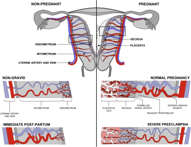 | 614 × 472 (143 KB) | S8600021 (talk | contribs) | Diagrammatic representation of uterine and placental vasculature (red shading = arterial; blue shading = venous) in the non-pregnant, pregnant and immediate post-partum state. Normal pregnancy is characterized by the formation of large arterio-venous sh |
You cannot overwrite this file.
File usage
The following 12 pages use this file: