File:Ureteral duplication 01.jpg

Original file (600 × 812 pixels, file size: 61 KB, MIME type: image/jpeg)
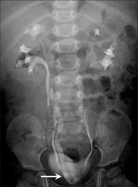
Ureteral Duplication
- Frontal radiograph from an intravenous urogram study shows bilateral complete ureteral duplication.
- The opening of the right upper moiety ureter is medial to the lower moiety ureter; there is ureterocele formation.
Reference
Alorainy IA, Barlas NB & Al-Boukai AA. (2010). Pictorial Essay: Infants of diabetic mothers. Indian J Radiol Imaging , 20, 174-81. PMID: 21042439 DOI.
Copyright
This is an open-access article distributed under the terms of the Creative Commons Attribution License, which permits unrestricted use, distribution, and reproduction in any medium, provided the original work is properly cited.
Original file name: Figure 10 IJRI-20-174-g010.jpg http://www.ncbi.nlm.nih.gov/pmc/articles/PMC2963757/figure/F0010/
Cite this page: Hill, M.A. (2024, May 21) Embryology Ureteral duplication 01.jpg. Retrieved from https://embryology.med.unsw.edu.au/embryology/index.php/File:Ureteral_duplication_01.jpg
- © Dr Mark Hill 2024, UNSW Embryology ISBN: 978 0 7334 2609 4 - UNSW CRICOS Provider Code No. 00098G
File history
Click on a date/time to view the file as it appeared at that time.
| Date/Time | Thumbnail | Dimensions | User | Comment | |
|---|---|---|---|---|---|
| current | 22:29, 2 May 2011 | | 600 × 812 (61 KB) | S8600021 (talk | contribs) | ==Ureteral duplication== Ureteral duplication: Frontal radiograph from an intravenous urogram study shows bilateral complete ureteral duplication. The opening of the right upper moiety ureter is medial to the lower moiety ureter; there is ureterocele for |
You cannot overwrite this file.
File usage
The following 2 pages use this file: