File:Trophoblast hCG function.jpg
Original file (600 × 1,798 pixels, file size: 229 KB, MIME type: image/jpeg)
Trophoblast hCG function
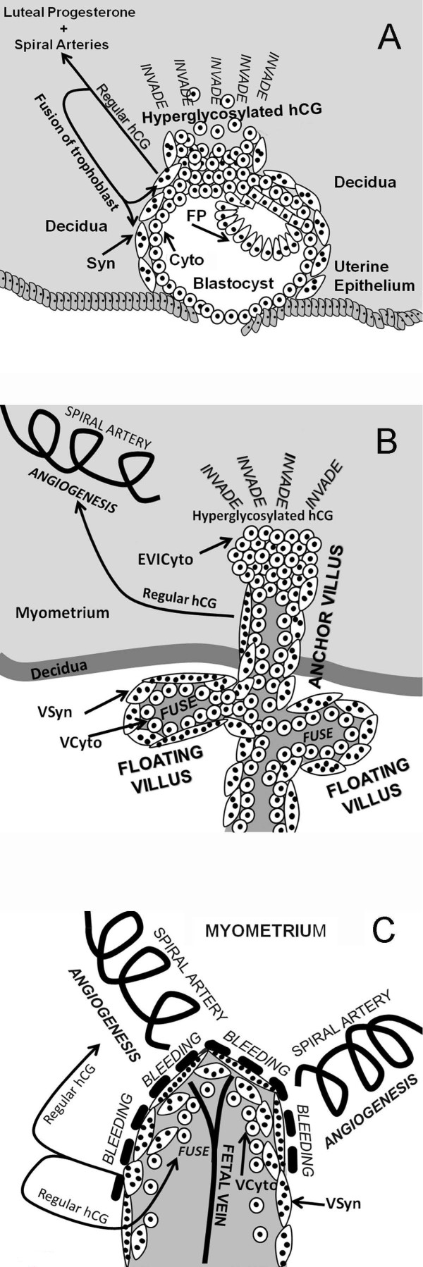
Villous placental tissue, cytotrophoblast and syncytiotrophoblast cells, and regular hCG and hyperglycosylated hCG function.
Panel A - illustrated blastocyst implantation and engulfment and trophoblast invasion at 3–5 weeks of gestation. Arrows illustrates the biological functions of regular hCG and hyperglycosylated hCG. Mononuclear cells are cytotrophoblast cells, cells with multiple nuclei (black circles) represen syncytiotrophoblast cells.
Panel B - villous trophoblast formation and function are illustrated at 6–8 weeks of gestation. Figure illustrates villous trophoblast (anchoring villus and floating villus) growth 5 to 10 weeks of gestation, and invasion of the decidua and myometrium in establishing hemochorial placentation.
Panel C - illustrates functional hemochorial placentation in floating villus at 10–12 weeks of gestation after invasion is complete. Cells with varying numbers of multiple nuclei (black circles) represent villous syncytiotrophoblast (VSyn), mononuclear cells are villous cytotrophoblast (VCyto) and extravillous invasive cytotrophoblast (EVICyto). Arrows shows biological actions of regular hCG and hyperglycosylated hCG.
Reference
Cole LA. (2009). New discoveries on the biology and detection of human chorionic gonadotropin. Reprod. Biol. Endocrinol. , 7, 8. PMID: 19171054 DOI.
Copyright
This is an Open Access article distributed under the terms of the Creative Commons Attribution License (http://creativecommons.org/licenses/by/2.0), which permits unrestricted use, distribution, and reproduction in any medium, provided the original work is properly cited. Copyright © 2009 Cole; licensee BioMed Central Ltd.
Reprod Biol Endocrinol. 2009; 7: 8. Published online 2009 January 26. doi: 10.1186/1477-7827-7-8.
Cite this page: Hill, M.A. (2024, April 27) Embryology Trophoblast hCG function.jpg. Retrieved from https://embryology.med.unsw.edu.au/embryology/index.php/File:Trophoblast_hCG_function.jpg
- © Dr Mark Hill 2024, UNSW Embryology ISBN: 978 0 7334 2609 4 - UNSW CRICOS Provider Code No. 00098G
File history
Click on a date/time to view the file as it appeared at that time.
| Date/Time | Thumbnail | Dimensions | User | Comment | |
|---|---|---|---|---|---|
| current | 20:52, 5 October 2009 | 600 × 1,798 (229 KB) | S8600021 (talk | contribs) | Villous placental tissue, cytotrophoblast and syncytiotrophoblast cells, and regular hCG and hyperglycosylated hCG function. Panel A illustrated blastocyst implantation and engulfment and trophoblast invasion at 3–5 weeks of gestation. Arrows illustrate |
You cannot overwrite this file.