File:Stage7 bf5.jpg: Difference between revisions
No edit summary |
mNo edit summary |
||
| Line 2: | Line 2: | ||
Appearance of section looks to be about a [[Carnegie stage 7]] embryo. | Appearance of section looks to be about a [[Carnegie stage 7]] embryo. | ||
=== Human Embryo Stage 7 Information=== | === Human Embryo Stage 7 Information=== | ||
| Line 21: | Line 17: | ||
Mesoderm lies between the ectoderm and endoderm as a continuous sheet except at the buccopharyngeal and cloacal membranes. These membranes have ectoderm and endoderm only and will lie at the rostral (head) and caudal (tail) of the gastrointestinal tract. | Mesoderm lies between the ectoderm and endoderm as a continuous sheet except at the buccopharyngeal and cloacal membranes. These membranes have ectoderm and endoderm only and will lie at the rostral (head) and caudal (tail) of the gastrointestinal tract. | ||
{{Stage7 bf5 links}} | |||
Original | :'''Image Links:''' [[:File:Stage7 bf5.jpg|Original full image]] | [[:File:Stage7 bf51.jpg|Trilaminar embryo excerpt]] | ||
:'''Image version links:''' [[:File:Stage7 bf5.jpg|ExtraLarge 1712x1206px]] | [[:File:Stage7 bf5a.jpg|Large 1024x721px]] | [[:File:Stage7 bf5b.jpg|Medium 500x352px]] | [[:File:Stage7 bf5c.jpg|Small 240x169px]] | |||
{{Ed Uthman}} | |||
[[Category:Human Embryo]] [[Category:Carnegie Stage]] [[Category:Carnegie Stage 7]] [[Category:Week 3]] [[Category:Gastrulation]] [[Category:Ectopic Pregnancy]] | [[Category:Human Embryo]] [[Category:Carnegie Stage]] [[Category:Carnegie Stage 7]] [[Category:Week 3]] [[Category:Gastrulation]] [[Category:Ectopic Pregnancy]] | ||
Revision as of 11:25, 3 March 2014
Human Embryo Stage 7
Appearance of section looks to be about a Carnegie stage 7 embryo.
Human Embryo Stage 7 Information
Carnegie stage 7
Features: embryonic disc, primitive node, primative streak, primitive groove, yolk sac
Facts: Week 3, 15 - 17 days, 0.4 mm
View 1: embryonic disc, showing the epiblast viewed from the amniotic (dorsal) side.
Events: Gastrulation is continuing as cells migrate from the epiblast, continuing to form mesoderm.
Mesoderm lies between the ectoderm and endoderm as a continuous sheet except at the buccopharyngeal and cloacal membranes. These membranes have ectoderm and endoderm only and will lie at the rostral (head) and caudal (tail) of the gastrointestinal tract.
- Stage 7 Links: Large image | Medium image | Small image | Trilaminar embryo excerpt | Villi excerpt 1 | Villi excerpt 2 | Carnegie stage 7
- Carnegie Stages: 1 | 2 | 3 | 4 | 5 | 6 | 7 | 8 | 9 | 10 | 11 | 12 | 13 | 14 | 15 | 16 | 17 | 18 | 19 | 20 | 21 | 22 | 23 | About Stages | Timeline
Image: Dr Ed Uthman (Houston, Texas) - other pathology images - CC BY 2.0
Cite this page: Hill, M.A. (2024, May 17) Embryology Stage7 bf5.jpg. Retrieved from https://embryology.med.unsw.edu.au/embryology/index.php/File:Stage7_bf5.jpg
- © Dr Mark Hill 2024, UNSW Embryology ISBN: 978 0 7334 2609 4 - UNSW CRICOS Provider Code No. 00098G
- Image Links: Original full image | Trilaminar embryo excerpt
- Image version links: ExtraLarge 1712x1206px | Large 1024x721px | Medium 500x352px | Small 240x169px
Image: Dr Ed Uthman (Houston, Texas) - other pathology images
File history
Click on a date/time to view the file as it appeared at that time.
| Date/Time | Thumbnail | Dimensions | User | Comment | |
|---|---|---|---|---|---|
| current | 14:43, 21 July 2010 | 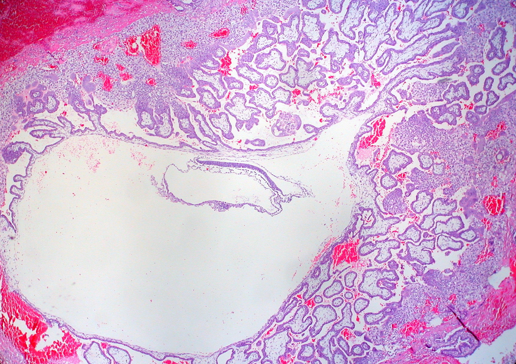 | 1,712 × 1,206 (875 KB) | S8600021 (talk | contribs) | ==Human embryo - 3 week== Appearance of section looks to be about a Carnegie stage 7 embryo. Original Author Legend - Primitive Trilaminar Human Embryo in Tubal Pregnancy (40X) :"I think this is at about the same developmental stage as the Hertig-Roc |
You cannot overwrite this file.
File usage
The following 9 pages use this file: