File:Seminiferous-tubule-HEx40.jpg: Difference between revisions
From Embryology
No edit summary |
mNo edit summary |
||
| (14 intermediate revisions by 2 users not shown) | |||
| Line 1: | Line 1: | ||
==Testis Histology - Seminiferous | ==Testis Histology - Seminiferous Tubule== | ||
[[File:Seminiferous tubule cartoon.jpg|thumb|Seminiferous tubule cartoon]] | |||
Seminiferous tubule cross-section {{HE}} (x40) | |||
* '''Smooth muscle''' - covers the outside of the seminiferous tubule. | |||
* '''{{Spermatogonia}}''' - arranged at the periphery of the seminiferous tubule. These cells are the diploid germ cell population that will undergo mitosis and then meiosis. | |||
* '''{{Sertoli cell}}s''' - arranged across all layers of the seminiferous tubule. These cells are the supporting cells, nutritional and mechanical, as well as forming a blood-testis barrier. | |||
* '''Primary spermatocytes''' - arranged deep (luminal) to the spermatogonia. These large cells enter the prophase of the first meiotic division. [[P#primary spermatocyte|primary spermatocyte]] | |||
* '''Secondary spermatocytes''' - small, complete the second meiotic division | |||
* '''Spermatid''' - lie deep (luminal) to the secondary spermatocyte. These small cells are haploid and in will undergo spermiogenesis, changing their cellular structure and shape to form spermatozoa. | |||
* '''Spermatozoa''' - differentiated gamete. | |||
===Sertoli cells=== | |||
* The {{Sertoli cell}} provides mechanical and nutritive support for the spermatogenic cells. | |||
* Secrete two hormones - inhibin and activin - which provide positive and negative feedback on FSH secretion from the pituitary. | |||
** activins - dimers of beta-A and/or beta-B subunits encoded by the genes INHBA and INHBB | |||
** Follicle-stimulating hormone (FSH)-releasing protein (FRP) subunit is identical in structure to the beta-A subunit of inhibin. | |||
* less numerous than the spermatogenic cells and are evenly distributed between them. | |||
* cell shape is highly irregular - columnar is the best approximation. | |||
* cells extend from the basement membrane to the luminal surface of the seminiferous epithelium. | |||
* processes of the Sertoli cells extend in between the spermatogenic cells (cell limits are therefore not clearly visible in the LM). | |||
* nucleus of Sertoli cells is ovoid or angular, large and lightly stained and often contains a large nucleolus. | |||
** long axis of the nucleus is oriented perpendicular to wall of the tubule. | |||
** a fold in the nuclear membrane is characteristic for Sertoli cells but not always visible. | |||
* lateral processes of Sertoli cells are interconnected by tight junctions, which are likely to be the structural basis for the blood-testis barrier. | |||
* Spermatogonia and primary spermatocytes are located in the basal compartment, other cellular stages of spermatogenesis are located in the adluminal compartment. | |||
* Tight junctions may temporarily open to permit the passage of spermatogenic cells from the basal into the adluminal compartment. | |||
---- | |||
{{Testis Histology}} | |||
{{Footer}} | |||
[[Category:Testis]] [[Category:Genital]] [[Category:Histology]] | [[Category:Testis]] [[Category:Genital]] [[Category:Histology]] | ||
Latest revision as of 10:37, 2 May 2019
Testis Histology - Seminiferous Tubule

Seminiferous tubule cross-section (Stain - Haematoxylin Eosin) (x40)
- Smooth muscle - covers the outside of the seminiferous tubule.
- spermatogonia - arranged at the periphery of the seminiferous tubule. These cells are the diploid germ cell population that will undergo mitosis and then meiosis.
- sertoli cells - arranged across all layers of the seminiferous tubule. These cells are the supporting cells, nutritional and mechanical, as well as forming a blood-testis barrier.
- Primary spermatocytes - arranged deep (luminal) to the spermatogonia. These large cells enter the prophase of the first meiotic division. primary spermatocyte
- Secondary spermatocytes - small, complete the second meiotic division
- Spermatid - lie deep (luminal) to the secondary spermatocyte. These small cells are haploid and in will undergo spermiogenesis, changing their cellular structure and shape to form spermatozoa.
- Spermatozoa - differentiated gamete.
Sertoli cells
- The sertoli cell provides mechanical and nutritive support for the spermatogenic cells.
- Secrete two hormones - inhibin and activin - which provide positive and negative feedback on FSH secretion from the pituitary.
- activins - dimers of beta-A and/or beta-B subunits encoded by the genes INHBA and INHBB
- Follicle-stimulating hormone (FSH)-releasing protein (FRP) subunit is identical in structure to the beta-A subunit of inhibin.
- less numerous than the spermatogenic cells and are evenly distributed between them.
- cell shape is highly irregular - columnar is the best approximation.
- cells extend from the basement membrane to the luminal surface of the seminiferous epithelium.
- processes of the Sertoli cells extend in between the spermatogenic cells (cell limits are therefore not clearly visible in the LM).
- nucleus of Sertoli cells is ovoid or angular, large and lightly stained and often contains a large nucleolus.
- long axis of the nucleus is oriented perpendicular to wall of the tubule.
- a fold in the nuclear membrane is characteristic for Sertoli cells but not always visible.
- lateral processes of Sertoli cells are interconnected by tight junctions, which are likely to be the structural basis for the blood-testis barrier.
- Spermatogonia and primary spermatocytes are located in the basal compartment, other cellular stages of spermatogenesis are located in the adluminal compartment.
- Tight junctions may temporarily open to permit the passage of spermatogenic cells from the basal into the adluminal compartment.
Testis Histology Links: Testis Development | Spermatozoa Development | Histology
- Human (young): overview labeled | overview unlabeled | convoluted seminiferous tubules x10 | x40 | x40 | tunica albuginea x20
- Human (adult): overview x2 | convoluted seminiferous tubules labeled | x10 | x20 | x40 | x40 | epididymis ductulus efferens | ductus epididymidis | epithelium | overview x4 | x10 | x20 | x40 | ductus deferens labeled overview | epithelium | overview x2 | x10 | x40
- Human Stage 22: Testis - labeled overview | Testis - unlabeled overview | Testis - unlabeled detail | Testis - labeled detail | testis | Carnegie stage 22 | Movie - Urogenital stage 22
- Mouse: postnatal epididymis | 14 days postnatal | 33 days postnatal | 45 days postnatal | 2 months postnatal
| Spermatozoa Development (expand to see terms) | ||
|---|---|---|
|
Note there are additional glossaries associated with genital, spermatozoa, oocyte and renal.
See also: Spermatozoa Terms collapse table
|

Cite this page: Hill, M.A. (2024, May 21) Embryology Seminiferous-tubule-HEx40.jpg. Retrieved from https://embryology.med.unsw.edu.au/embryology/index.php/File:Seminiferous-tubule-HEx40.jpg
- © Dr Mark Hill 2024, UNSW Embryology ISBN: 978 0 7334 2609 4 - UNSW CRICOS Provider Code No. 00098G
File history
Click on a date/time to view the file as it appeared at that time.
| Date/Time | Thumbnail | Dimensions | User | Comment | |
|---|---|---|---|---|---|
| current | 09:16, 3 May 2010 | 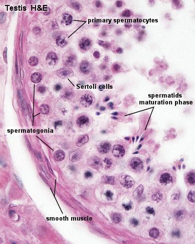 | 400 × 500 (59 KB) | S8600021 (talk | contribs) | Seminiferous tubule (HEx40) Category:Testes Category:Genital Category:Histology |
You cannot overwrite this file.
File usage
The following 20 pages use this file:
- 2010 BGD Lecture - Development of the Embryo/Fetus 1
- 2010 BGD Practical 3 - Gametogenesis
- 2011 Lab 1 - Gametogenesis
- 2011 Lab 1 - Spermatogenesis
- ANAT2241 Male Reproductive System
- ANAT2341 Lab 1 - Gametogenesis
- ANAT2341 Lab 1 - Spermatogenesis
- BGDA Lecture - Development of the Embryo/Fetus 1
- BGDA Practical - Male Reproductive Tract Histology
- BGDA Practical 3 - Gametogenesis
- BGD Lecture - Sexual Differentiation
- Cell Division - Meiosis
- Lecture - Fertilization
- Lecture - Genital Development
- Sertoli cell
- Spermatozoa Development
- Testis Development
- Talk:BGDA Practical 3 - Gametogenesis
- User:Z3418779
- File:Seminiferous tubule cartoon.jpg