File:Respiratory histology 14.jpg: Difference between revisions
From Embryology
mNo edit summary |
mNo edit summary |
||
| Line 16: | Line 16: | ||
{{Nasal olfactory links}} | |||
Latest revision as of 12:36, 10 March 2013
Nasal Cavity Olfactory Region Histology
Olfactory epithelium cells
- Olfactory cells
- Sustentacular cells - located mainly in the superficial cell layer of the epithelium (difficult to distinguish from olfactory cells).
- Basal cells - identified by their location.
Epithelium
- Cilia are not visible
- goblet cells are absent from the olfactory epithelium.
Lamina Propria
- olfactory axon bundles (lightly stained, rounded areas) connected to olfactory cells.
- Bowman's glands - (small mucous glands, olfactory glands) function to moisturise the epithelium.
- Nasal Olfactory Histology: overview image | detail image | Smell Development | Histology | Histology Stains
- Respiratory Histology: Bronchiole | Alveolar Duct | Alveoli | EM Alveoli septum | Alveoli Elastin | Trachea 1 | Trachea 2 | labeled lung | unlabeled lung | Respiratory Bronchiole | Lung Reticular Fibres | Nasal Inferior Concha | Nasal Respiratory Epithelium | Olfactory Region overview | Olfactory Region Epithelium | Histology Stains
File history
Click on a date/time to view the file as it appeared at that time.
| Date/Time | Thumbnail | Dimensions | User | Comment | |
|---|---|---|---|---|---|
| current | 23:06, 28 February 2012 | 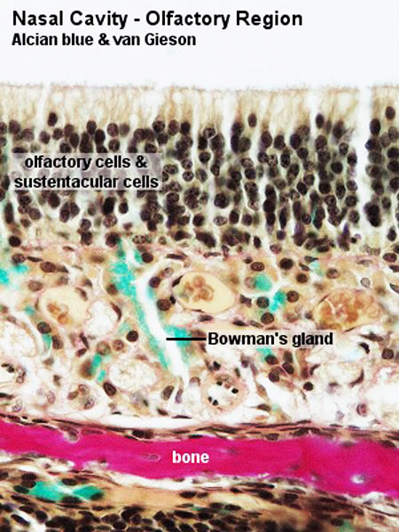 | 450 × 600 (87 KB) | Z8600021 (talk | contribs) |
You cannot overwrite this file.
File usage
The following 10 pages use this file: