File:Placenta humans and guinea-pig cartoon.jpg
Original file (1,200 × 889 pixels, file size: 562 KB, MIME type: image/jpeg)
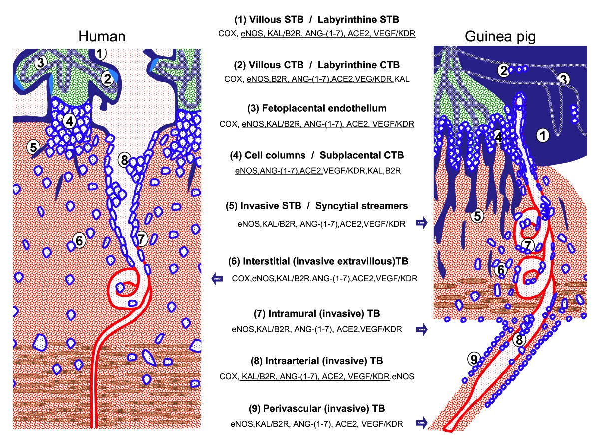
placenta and fetoplacental junctional zone of humans and guinea-pigs
Schematic representation of the placenta and fetoplacental junctional zone of humans and guinea-pigs. In order to highlight comparable structures, all fetal tissues are colored blue and green, all maternal structures red and brown. The main cellular and syncytial structures of both placentas are listed together with the reported expression of the vasodilator factors and enzymes. Factors are underlined when present in both species; isolated factors expressed only in one of the two species are lateralized to the presenting species. When the whole panel of factors is described in one of the species, this is depicted by a blue arrow pointing to the respective structures. Note that in the human an equivalent for periarterial trophoblast is not known. TB = trophoblasts; CTB = cytotrophoblasts; STB = syncytiotrophoblast.
Reference
Valdes et al. Reproductive Biology and Endocrinology 2009 7:79 doi:10.1186/1477-7827-7-79 1477-7827-7-79-8-l.jpg
File history
Click on a date/time to view the file as it appeared at that time.
| Date/Time | Thumbnail | Dimensions | User | Comment | |
|---|---|---|---|---|---|
| current | 11:21, 12 May 2013 | | 1,200 × 889 (562 KB) | Z8600021 (talk | contribs) | ==placenta and fetoplacental junctional zone of humans and guinea-pigs== Schematic representation of the placenta and fetoplacental junctional zone of humans and guinea-pigs. In order to highlight comparable structures, all fetal tissues are colored b... |
You cannot overwrite this file.
File usage
The following page uses this file: