File:Placenta and explant model.jpg
Original file (800 × 682 pixels, file size: 153 KB, MIME type: image/jpeg)
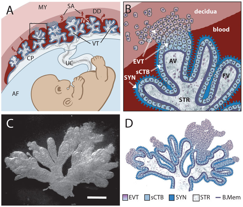
Comparison of in vivo placental structure to placenta explant model.
(A) Structure and orientation of fetus and placenta in uterus at ~6 weeks of gestation. Fetal structures are represented in shades of blue and purple while maternal are in shades of red. Maternal structures: MY: myometrium, SA: spiral arteries, DD: decidua (uterine lining during pregnancy), IVS: intervillous space filled with maternal blood. Fetal structures: VT: villous tree, CP: chorionic plate, UC: umbilical cord, AF: amniotic fluid.
(B) (Enlargement of boxed area in panel A) Maternal blood surrounds the villous tree composed of anchoring (AV) and floating villi (FV), which are covered by a syncytiotrophoblast (SYN) that is underlaid by subsyncytial cytotrophoblasts (sCTB) and a basement membrane. The subsyncytial CTB layer grows increasingly discontinuous in later trimesters. Gas and nutrient exchange with the maternal blood occurs across the syncytiotrophoblast to supply fetal capillaries in the stroma (STR). At the uterine wall, extravillous cytotrophoblasts (EVT) anchor the villous tree in the decidua. Some invade the decidua and move away from the tip to remodel maternal spiral arteries, with altered gene expression patterns as they move (not shown). Notably, E-cadherin expression decreases as VE-cadherin expression rises in distal (relative to fetus) extravillous cytotrophoblasts.
(C) A six-week placental explant anchored in Matrigel. Bar = 1 mm.
(D) Cartoon representation of the relevant structures seen in panel C.
Journal.ppat.1000732.g001.jpg
File history
Click on a date/time to view the file as it appeared at that time.
| Date/Time | Thumbnail | Dimensions | User | Comment | |
|---|---|---|---|---|---|
| current | 23:59, 21 April 2010 | | 800 × 682 (153 KB) | S8600021 (talk | contribs) | Comparison of in vivo placental structure to placenta explant model. (A) Structure and orientation of fetus and placenta in uterus at ~6 weeks of gestation. Fetal structures are represented in shades of blue and purple while maternal are in shades of red |
You cannot overwrite this file.