File:Placenta Hofbauer cells 01.jpg

Original file (934 × 700 pixels, file size: 156 KB, MIME type: image/jpeg)
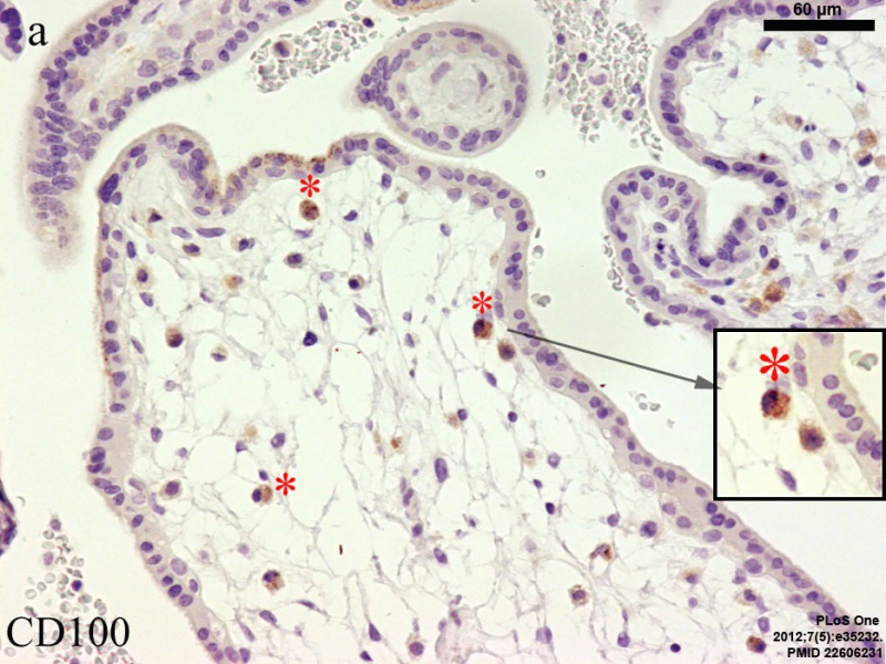
Placenta Hofbauer cells
Tissue from a representative first miscarriage, 9th week of gestation (GA week 9; approx. post-fertilization age week 7).
Paraffin serial sections. A subpopulation of Hofbauer cells residing in the villous stroma, identified by are positive for CD100 (panel a, red asterisks)
Bar = 60 µm.
Reference
<pubmed>22606231</pubmed>| PLoS One.
Copyright: © 2012 Lorenzi et al. This is an open-access article distributed under the terms of the Creative Commons Attribution License, which permits unrestricted use, distribution, and reproduction in any medium, provided the original author and source are credited.
Figure 2. panel A resized and contrast adjusted. doi:10.1371/journal.pone.0035232.g002 Journal.pone.0035232.g002.jpg
File history
Click on a date/time to view the file as it appeared at that time.
| Date/Time | Thumbnail | Dimensions | User | Comment | |
|---|---|---|---|---|---|
| current | 13:51, 20 November 2012 | | 934 × 700 (156 KB) | Z8600021 (talk | contribs) | |
| 13:35, 20 November 2012 | 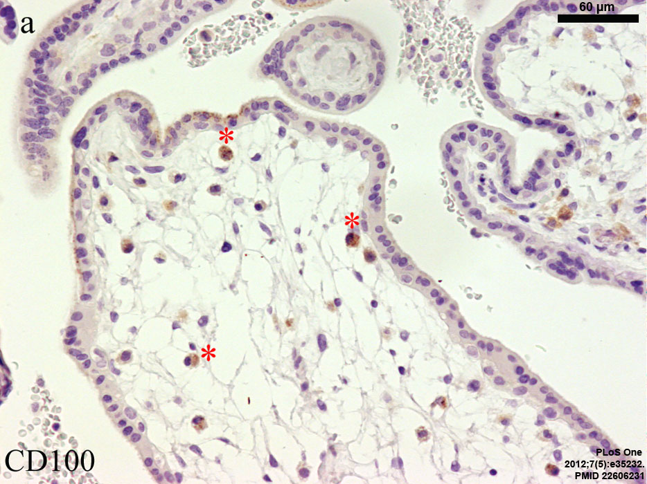 | 934 × 700 (153 KB) | Z8600021 (talk | contribs) | ==Placenta Hofbauer cells== Figure 2. Tissue from a representative first miscarriage, 9th week of gestation. Paraffin serial sections. A subpopulation of Hofbauer cells residing in the villous stroma, identified by are positive for CD100 (panel a, red a |
You cannot overwrite this file.
File usage
The following 4 pages use this file: