File:Paramesonephric duct.jpg
Paramesonephric_duct.jpg (423 × 478 pixels, file size: 40 KB, MIME type: image/jpeg)
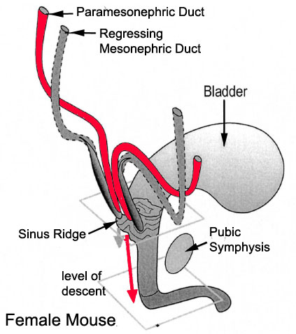
Paramesonephric Duct
Data from the developing mouse showing the relationship between the mesonephric and paramesonephric ducts opening into the urogenital sinus.
The paramesonephric duct began as an infold of surface epithelium lying along the surface of the genital ridge. Estrogens, both maternal and fetal, stimulate its development and that eventually of the external female fetal genital structures.
In contrast, the mesonephric duct regresses, remnants of this duct may remain lying within the broad ligament.
Image modified from: Drews U, Sulak O, Schenck PA. Androgens and the development of the vagina. Biol Reprod. 2002 Oct;67(4):1353-9. PMID: 12297555
File history
Click on a date/time to view the file as it appeared at that time.
| Date/Time | Thumbnail | Dimensions | User | Comment | |
|---|---|---|---|---|---|
| current | 22:49, 21 September 2009 | | 423 × 478 (40 KB) | S8600021 (talk | contribs) |
You cannot overwrite this file.
File usage
The following 13 pages use this file:
- 2009 Lecture 16
- 2010 Lecture 16
- 2011 Lab 8 - Fetal
- 2011 Lecture 16
- 2014 Group Project 4
- ANAT2341 Lab 8 - Fetal
- BGDB Sexual Differentiation - Fetal
- BGD Lecture - Sexual Differentiation
- Lecture - Genital Development
- REI - Reproductive Medicine Seminar 2018
- Royal Hospital for Women - Reproductive Medicine Seminar 2018
- Uterus Development
- Talk:2014 Group Project 4