File:Nail structure cartoon1.jpg
Nail_structure_cartoon1.jpg (500 × 377 pixels, file size: 54 KB, MIME type: image/jpeg)
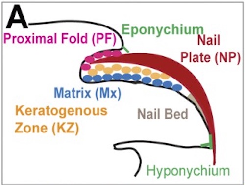
Mouse Nail Components
Reference
Leung Y, Kandyba E, Chen YB, Ruffins S, Chuong CM & Kobielak K. (2014). Bifunctional ectodermal stem cells around the nail display dual fate homeostasis and adaptive wounding response toward nail regeneration. Proc. Natl. Acad. Sci. U.S.A. , 111, 15114-9. PMID: 25277970 DOI.
Copyright
Proceedings National Academy of Sciences (PNAS) Liberalization of PNAS copyright policy: Noncommercial use freely allowed Note original Author should be contacted for permission to reuse for Educational purposes. See also PNAS Author Rights and Permission FAQs
- Cozzarelli NR, Fulton KR, Sullenberger DM. Liberalization of PNAS copyright policy: noncommercial use freely allowed. Proc Natl Acad Sci U S A. 2004 Aug 24;101(34):12399. PMID15314225 "Our guiding principle is that, while PNAS retains copyright, anyone can make noncommercial use of work in PNAS without asking our permission, provided that the original source is cited."
Panel A cropped from full figure - Pnas.1318848111fig01.jpg
Cite this page: Hill, M.A. (2024, May 21) Embryology Nail structure cartoon1.jpg. Retrieved from https://embryology.med.unsw.edu.au/embryology/index.php/File:Nail_structure_cartoon1.jpg
- © Dr Mark Hill 2024, UNSW Embryology ISBN: 978 0 7334 2609 4 - UNSW CRICOS Provider Code No. 00098G
File history
Click on a date/time to view the file as it appeared at that time.
| Date/Time | Thumbnail | Dimensions | User | Comment | |
|---|---|---|---|---|---|
| current | 12:29, 13 January 2019 | | 500 × 377 (54 KB) | Z8600021 (talk | contribs) |
You cannot overwrite this file.
File usage
The following page uses this file: