File:Mouse neonatal ovary oocyte EM02.jpg
From Embryology
Size of this preview: 598 × 600 pixels. Other resolution: 790 × 792 pixels.
Original file (790 × 792 pixels, file size: 179 KB, MIME type: image/jpeg)
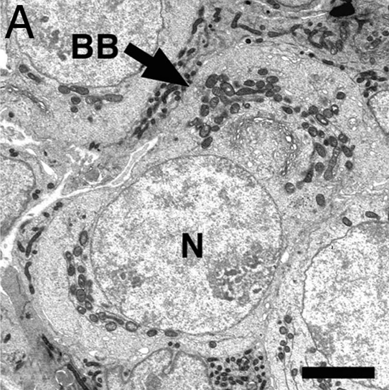
Electron Micrographs of Oocytes in Neonatal Mouse Ovaries
- A - Micrograph of an oocyte within a germline cyst from PND1 showing a well defined Balbiani body (arrow) with Golgi surrounded by mitochondria.
- EM Links: All Images | balbini_body | A | B | C | D | E | F | Oocyte Development | Ovary Development | Mouse Development
Legend
- BB - Balbiani body
- M - mitochondria
- n - nucleus
Scale bar: 5 microns
Reference
<pubmed>17189423</pubmed>| PNAS
Copyright
Proceedings National Academy of Sciences (PNAS) Liberalization of PNAS copyright policy: Noncommercial use freely allowed Note original Author should be contacted for permission to reuse for Educational purposes. See also PNAS Author Rights and Permission FAQs
- Cozzarelli NR, Fulton KR, Sullenberger DM. Liberalization of PNAS copyright policy: noncommercial use freely allowed. Proc Natl Acad Sci U S A. 2004 Aug 24;101(34):12399. PMID15314225 "Our guiding principle is that, while PNAS retains copyright, anyone can make noncommercial use of work in PNAS without asking our permission, provided that the original source is cited."
Mouse neonatal ovary oocyte EM01.jpg
File history
Click on a date/time to view the file as it appeared at that time.
| Date/Time | Thumbnail | Dimensions | User | Comment | |
|---|---|---|---|---|---|
| current | 18:32, 2 March 2012 | | 790 × 792 (179 KB) | Z8600021 (talk | contribs) | ==Electron Micrographs of Oocytes in Neonatal Mouse Ovaries== * '''A''' - Micrograph of an oocyte within a germline cyst from PND1 showing a well defined Balbiani body (arrow) with Golgi surrounded by mitochondria. ===Legend=== * '''BB''' - Balbiani |
You cannot overwrite this file.
File usage
There are no pages that use this file.