File:Mouse germinal vesicle 01.jpg
Original file (1,200 × 599 pixels, file size: 106 KB, MIME type: image/jpeg)
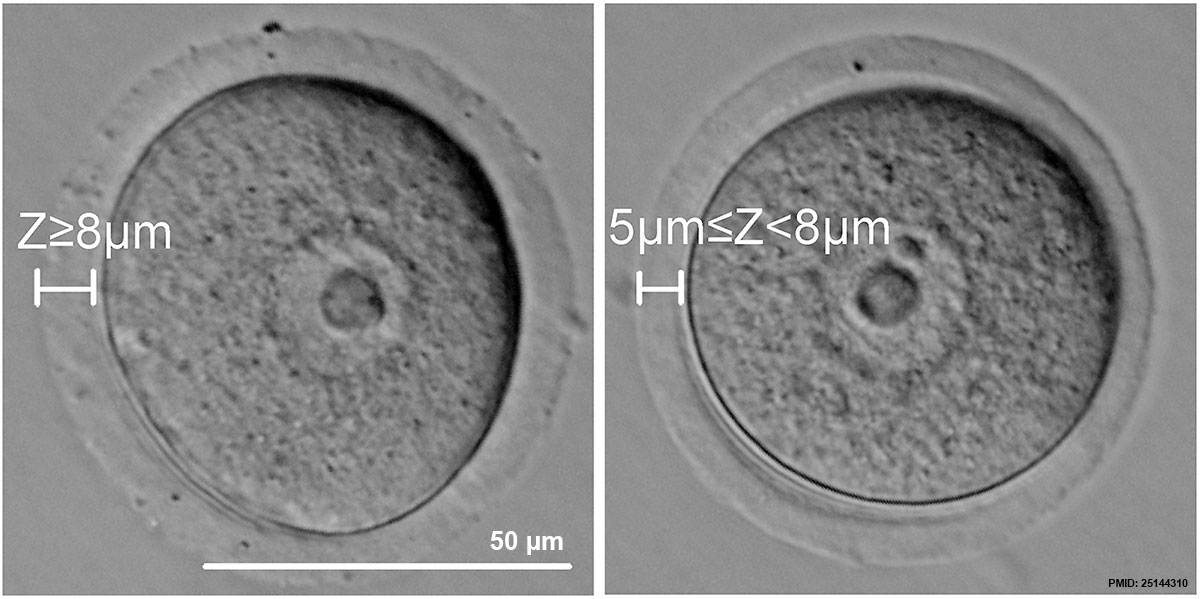
GV stage oocyte classification based on thickness of the ZP
GV stage COCs were collected from ovaries from mice following administration of PMSG. The COCs were classified into three categories according to the thickness of their ZP (Z): COCs with Z≥8 µm (upper line, left), COCs with 5 µm≤Z<8 µm (upper line, middle), COCs with Z<5 µm (upper line, right). For the ZP digestion experiment, oocytes with Z≥8 µm (bottom line, left) were treated with tyrode solution to generate 5 µm≤Z<8 µm (bottom line, right). Scale = 50 µm.
- Mouse Germinal Vesicle Links: Image - 1 | Image - 2 | Image - 3 | Image - 4 | Oocyte Development | Zona pellucida | Granulosa cell | Mouse Development
Reference
<pubmed>25144310</pubmed>| PLoS One.
Copyright
© 2014 Zhou et al. This is an open-access article distributed under the terms of the Creative Commons Attribution License, which permits unrestricted use, distribution, and reproduction in any medium, provided the original author and source are credited.
PLoS One. 2014 Aug 21;9(8):e105812. doi: 10.1371/journal.pone.0105812. eCollection 2014. Assessment of mouse germinal vesicle stage oocyte quality by evaluating the cumulus layer, zona pellucida, and perivitelline space. Zhou HX1, Ma YZ2, Liu YL1, Chen Y1, Zhou CJ1, Wu SN1, Shen JP1, Liang CG1. Author information
Abstract To improve the outcome of assisted reproductive technology (ART) for patients with ovulation problems, it is necessary to retrieve and select germinal vesicle (GV) stage oocytes with high developmental potential. Oocytes with high developmental potential are characterized by their ability to undergo proper maturation, fertilization, and embryo development. In this study, we analyzed morphological traits of GV stage mouse oocytes, including cumulus cell layer thickness, zona pellucida thickness, and perivitelline space width. Then, we assessed the corresponding developmental potential of each of these oocytes and found that it varies across the range measured for each morphological trait. Furthermore, by manipulating these morphological traits in vitro, we were able to determine the influence of morphological variation on oocyte developmental potential. Manually altering the thickness of the cumulus layer showed strong effects on the fertilization and embryo development potentials of oocytes, whereas manipulation of zona pellucida thickness effected the oocyte maturation potential. Our results provide a systematic detailed method for selecting GV stage oocytes based on a morphological assessment approach that would benefit for several downstream ART applications.
PMID 25144310
Figure 2. doi:10.1371/journal.pone.0105812.g002
File history
Click on a date/time to view the file as it appeared at that time.
| Date/Time | Thumbnail | Dimensions | User | Comment | |
|---|---|---|---|---|---|
| current | 21:31, 8 September 2014 | | 1,200 × 599 (106 KB) | Z8600021 (talk | contribs) | ==GV stage oocyte classification based on thickness of the ZP== GV stage COCs were collected from ovaries from mice following administration of PMSG. The COCs were classified into three categories according to the thickness of their ZP (Z): COCs with... |
You cannot overwrite this file.
File usage
The following page uses this file: