File:Mouse cerebellar foliation defects.jpg
Mouse_cerebellar_foliation_defects.jpg (574 × 600 pixels, file size: 115 KB, MIME type: image/jpeg)
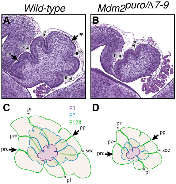
Mouse Cerebellar Foliation Defects
(A–B) Midsagittal sections of newborn (P0) wild-type and Mdm2puro/Δ7-9 cerebella stained with H&E. The four principal fissures, denoted by asterisks, are apparent in wild-type cerebella at P0 (A), whereas only two are evident in Mdm2puro/Δ7-9 mice (B). Wild-type pups show two additional fissures, indicated by arrows, whereas mutant pups do not. (C–D) Superimposition of P0 (purple outline), P7 (blue outline), and adult (green outline) cerebella from wild-type (C) or Mdm2puro/Δ7-9 (D) mice. By P7, all four primary fissures, as well as two additional fissures, are evident in Mdm2puro/Δ7-9 mice. Moreover, even in adulthood, the mutant cerebellum does not reach the size or complexity of the wild-type cerebellum. Abbreviations are: prc, precentral; pc, pre-culminate; pr, primary; pp, prepyramidal; sec, secondary; pl, posterolateral fissures.
Figure 3. Journal.pone.0017884.g003.jpg
Reference
<pubmed>21437245</pubmed>| PLoS One.
File history
Click on a date/time to view the file as it appeared at that time.
| Date/Time | Thumbnail | Dimensions | User | Comment | |
|---|---|---|---|---|---|
| current | 23:55, 22 September 2011 | | 574 × 600 (115 KB) | S8600021 (talk | contribs) | ==Mouse Cerebellar Foliation Defects== (A–B) Midsagittal sections of newborn (P0) wild-type and Mdm2puro/Δ7-9 cerebella stained with H&E. The four principal fissures, denoted by asterisks, are apparent in wild-type cerebella at P0 (A), whereas only tw |
You cannot overwrite this file.
File usage
The following page uses this file: