File:Keibel Mall 266.jpg
Original file (740 × 608 pixels, file size: 77 KB, MIME type: image/jpeg)
- Axial Skeleton: Fig. 231 to Fig. 273 | Vertebral Column and Thorax | Occipital Region | XI. Development of the Skeleton and of the Connective Tissues
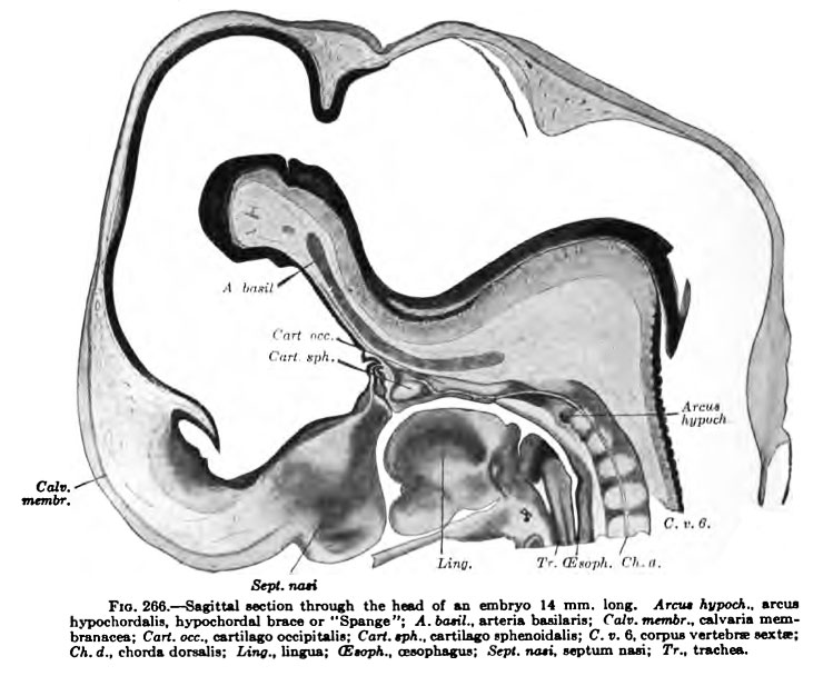
Meanwhile, during the period of chondrification in the arches and bodies of the cervical vertebrae, there takes place a condensation of tissue on the ventral margin of each of the more proximal cervical intervertebral discs near the cranial end of the vertebral body which lies caudalwards from it (Fig. 266). These condensed transverse bands of tissue connect the ventral ends of the blastemal neural processes with one another. They represent the hypochordal Spangen or braces of Froriep, and may appropriately be called hypochordal arches. In their intimate relations to the intervertebral discs they apparently differ from the hypochordal Spangen described by Charlotte Miiller in the thoracic region of the human embryo (see note, p. 334). In man the hypochordal arches are transitory in all except the first cervical segment. In the more distal segments the tissue composing them seems to become merged in the intervertebral discs without going beyond the blastemal stage. In the first cervical segment the hypochordal arch becomes chondrified at the time of the separation of the arches from the body after the temporary fusion mentioned above. The cartilage of the hypochordal arch becomes united on each side to that of the neural hemiarch. There are evidences of two bilaterally placed centres of chondrification in the hypochordal arch but fusion of these centres with one another and with the cartilage of the neural hemiarches takes place as soon as ehondrification is well under way. according to Froriep (1883), in the cow there is a single median centre in the hypochordal arch. In the white rat, according to Weiss, there are two bilaterally placed centres of chondrification in the hypochordal arch of the atlas.
- KM Figure Links: The Germ Cells | Segmentation | First Primitive Segment | Gastrulation | External Form | Placenta | Axial Skeleton | Limb Skeleton | Skull | Muscular System
| Historic Disclaimer - information about historic embryology pages |
|---|
 |
Glossary Links
- Glossary: A | B | C | D | E | F | G | H | I | J | K | L | M | N | O | P | Q | R | S | T | U | V | W | X | Y | Z | Numbers | Symbols | Term Link
Cite this page: Hill, M.A. (2024, May 21) Embryology Keibel Mall 266.jpg. Retrieved from https://embryology.med.unsw.edu.au/embryology/index.php/File:Keibel_Mall_266.jpg
- © Dr Mark Hill 2024, UNSW Embryology ISBN: 978 0 7334 2609 4 - UNSW CRICOS Provider Code No. 00098G
File history
Click on a date/time to view the file as it appeared at that time.
| Date/Time | Thumbnail | Dimensions | User | Comment | |
|---|---|---|---|---|---|
| current | 17:11, 2 October 2012 | | 740 × 608 (77 KB) | Z8600021 (talk | contribs) | {{KM Skeleton}} {{Keibel_Mall Images}} Category:Human Category:Bone Category:Axial Skeleton |
You cannot overwrite this file.
File usage
The following 2 pages use this file: