File:Integumentary histology 10.jpg

Original file (800 × 1,000 pixels, file size: 101 KB, MIME type: image/jpeg)
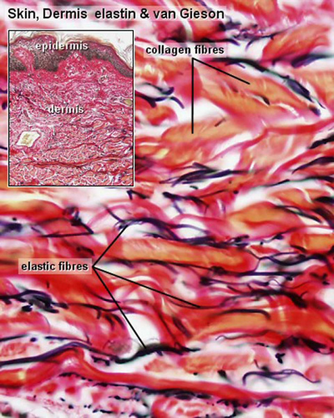
Integumentary Histology - Elastic Fibres
Skin, human - elastin & van Gieson
Elastic fibres require special stains to be visualised.
This histology image shows both elastic fibres and collagen bundles within the connective tissue of the dermis.
- Integument Histology Links: Adult Skin | Epidermis and Dermis | Thin Skin Epidermis | Thick Skin Epidermis | Elastic Fibres | Basal Cell Melanin | Foundations Practical Support | Integumentary System Development | Histology Stains
Links: Histology | Histology Stains | Blue Histology images copyright Lutz Slomianka 1998-2009. The literary and artistic works on the original Blue Histology website may be reproduced, adapted, published and distributed for non-commercial purposes. See also the page Histology Stains.
Cite this page: Hill, M.A. (2024, May 21) Embryology Integumentary histology 10.jpg. Retrieved from https://embryology.med.unsw.edu.au/embryology/index.php/File:Integumentary_histology_10.jpg
- © Dr Mark Hill 2024, UNSW Embryology ISBN: 978 0 7334 2609 4 - UNSW CRICOS Provider Code No. 00098G
Skk040evg.jpg
File history
Click on a date/time to view the file as it appeared at that time.
| Date/Time | Thumbnail | Dimensions | User | Comment | |
|---|---|---|---|---|---|
| current | 13:25, 9 March 2018 | | 800 × 1,000 (101 KB) | Z8600021 (talk | contribs) | ==Integumentary Histology - Elastic Fibres== Skin, human - elastin & van Gieson Elastic fibres require special stains to be visualised. {{Integument Histology}} {{Blue Histology}} Category:Integumentary Category:Histology Skk040evg.jpg |
You cannot overwrite this file.
File usage
The following page uses this file: