File:Ingalls1920plate05.jpg
From Embryology

Size of this preview: 454 × 599 pixels. Other resolution: 823 × 1,086 pixels.
Original file (823 × 1,086 pixels, file size: 160 KB, MIME type: image/jpeg)
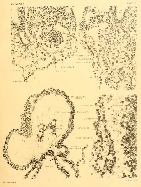
Plate 5
(Further details concerning the figures on this plate will be found in the text.)
Fig. 9.
- Section 12-1-2. X 350 Portion of body-stalk low down, showing blood-islands in left umbilical artery.
- Toward the yolk-sac, above in the illustration, the lumen of the artery communicates freely with the spaces in the mesenchyme.
Fig. 10.
- Section 12-1-6. X 350. Right umbilical artery in body-stalk, at a slightly higher level than figure 9.
- The vessel wall is here much better defined than that shown in figure 9.
- The blood-island i£ also less dense and almost free within the vessel.
- Toward the yolk-sac the changes in the vessel wall and relation of its lumen to the wide tissue spaces in front can be seen.
- This represents the first indication of a connection between the vessels in the embryo and those of the body-stalk; in this case between the right dorsal aorta and the right umbilical artery, through the intermediation of the caudal portion of the vitelline plexus and of the vitelline artery.
Fig. 11.
- Section 12-3-5. X 350. Detail of text-figure a. Section through heart, about the middle of the organ.
- The thickening of the mantle on the right side of the heart is only apparent, being due to the sudden change in curvature at this level.
Fig. 12.
- Section 12-2-6. X 600. Caudal end of left dorsal aorta, represented by an elongated blood island.
- Beginning lumen formation near its posterior end.
- The mesoderm in this region has not yet begun to segment.
- Embryo at Segmentation: Figure A | Plate 1 | Plate 2 | Plate 3 | Plate 4 | Plate 5 | Carnegie stage 9 | Carnegie Embryo 1878
Reference
Ingalls NW. A human embryo at the beginning of segmentation, with special reference to the vascular system. (1920) Contrib. Embryol., Carnegie Inst. Wash. Publ. 274, 11: 61-90.
Cite this page: Hill, M.A. (2024, April 27) Embryology Ingalls1920plate05.jpg. Retrieved from https://embryology.med.unsw.edu.au/embryology/index.php/File:Ingalls1920plate05.jpg
- © Dr Mark Hill 2024, UNSW Embryology ISBN: 978 0 7334 2609 4 - UNSW CRICOS Provider Code No. 00098G
| Historic Disclaimer - information about historic embryology pages |
|---|
| Pages where the terms "Historic" (textbooks, papers, people, recommendations) appear on this site, and sections within pages where this disclaimer appears, indicate that the content and scientific understanding are specific to the time of publication. This means that while some scientific descriptions are still accurate, the terminology and interpretation of the developmental mechanisms reflect the understanding at the time of original publication and those of the preceding periods, these terms, interpretations and recommendations may not reflect our current scientific understanding. (More? Embryology History | Historic Embryology Papers) |
File history
Click on a date/time to view the file as it appeared at that time.
| Date/Time | Thumbnail | Dimensions | User | Comment | |
|---|---|---|---|---|---|
| current | 18:21, 30 January 2012 |  | 823 × 1,086 (160 KB) | S8600021 (talk | contribs) | Reverted to version as of 08:08, 30 January 2012 |
| 18:17, 30 January 2012 |  | 938 × 1,192 (67 KB) | S8600021 (talk | contribs) | correct image | |
| 18:08, 30 January 2012 |  | 823 × 1,086 (160 KB) | S8600021 (talk | contribs) | {{Ingalls1920}} {{Historic Disclaimer}} |
You cannot overwrite this file.
File usage
The following 4 pages use this file:
