File:Ingalls1920plate03.jpg
From Embryology

Size of this preview: 497 × 599 pixels. Other resolution: 956 × 1,152 pixels.
Original file (956 × 1,152 pixels, file size: 70 KB, MIME type: image/jpeg)
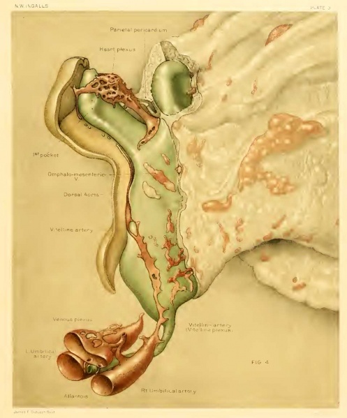
Plate 3
Fig. 4. Vascular system as seen from the right side
- X 100. Color scheme as in figure 3.
- The ectoderm has been cut away close to the border of the medullary folds.
- The mesoderm of the embryo and adjacent yolksac has been removed to show the vessels embedded in it.
- In the vitelline plexus and that part of it which represents the yolk-sac portion of the vitelline (umbilical) artery, the endothelial vessels are represented in darker color than the less definitely lined channels.
- The same is true for the anterior portion of the dorsal aorta and the isolated vesicles in the roof of the fore-gut.
- Scattered blood-islands and other vascular formations are seen through the mesoderm of the yolk-sac.
- Anterior to the branch of the aorta (marked vitelline artery) is a second, more doubtful connection between the aorta and the vitelline plexus.
- Embryo at Segmentation: Figure A | Plate 1 | Plate 2 | Plate 3 | Plate 4 | Plate 5 | Carnegie stage 9 | Carnegie Embryo 1878
Reference
Ingalls NW. A human embryo at the beginning of segmentation, with special reference to the vascular system. (1920) Contrib. Embryol., Carnegie Inst. Wash. Publ. 274, 11: 61-90.
Cite this page: Hill, M.A. (2024, April 27) Embryology Ingalls1920plate03.jpg. Retrieved from https://embryology.med.unsw.edu.au/embryology/index.php/File:Ingalls1920plate03.jpg
- © Dr Mark Hill 2024, UNSW Embryology ISBN: 978 0 7334 2609 4 - UNSW CRICOS Provider Code No. 00098G
| Historic Disclaimer - information about historic embryology pages |
|---|
 |
File history
Click on a date/time to view the file as it appeared at that time.
| Date/Time | Thumbnail | Dimensions | User | Comment | |
|---|---|---|---|---|---|
| current | 18:05, 30 January 2012 | | 956 × 1,152 (70 KB) | S8600021 (talk | contribs) | {{Ingalls1920}} {{Historic Disclaimer}} |
You cannot overwrite this file.
File usage
The following 4 pages use this file: