File:Ingalls1920FigureA.jpg

Original file (1,000 × 504 pixels, file size: 66 KB, MIME type: image/jpeg)
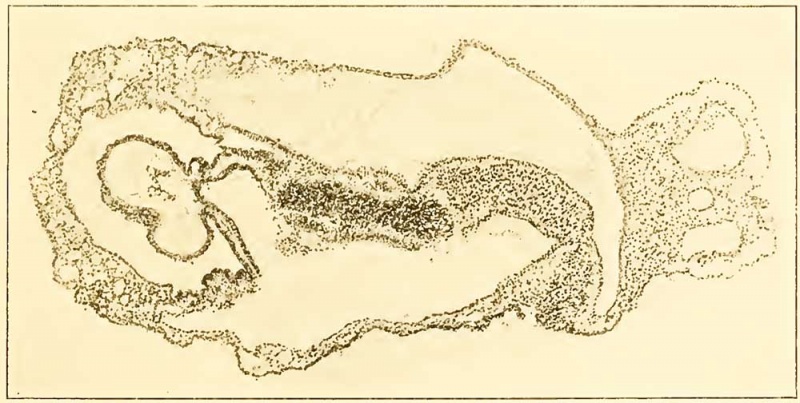
Figure A
Section 12-3-6, X90. The line of this section is shown in figure 3 on plate 2.
Transversely the plane of section is somewhat oblique, so that structures on the left of the median line (above in the illustration) are cut at a higher level than those on the right.
The section cuts just behind the primitive node, through the anterior intestinal portal and approximately, though obliquely, through the middle of the heart. On the right in the figure appears the body-stalk with the two umbilical veins and allantois between them.
Three cavities are seen: above in the illustration is the amniotic cavity; below is the yolk-sac, communicating anteriorly with the foregut; farther forward, in the median line, is the pericardial coelom with its contained heart.
The central, densely cellular mass is a tangential section of the left neural fold.
Between the entoderm of the foregut and the superficial ectoderm, the mesoderm can be traced forward to the point where it splits to become continuous with the visceral and parietal layers of the pericardial cavity. (Cf. also text and fig. 11, plate 5.)
- Embryo at Segmentation: Figure A | Plate 1 | Plate 2 | Plate 3 | Plate 4 | Plate 5 | Carnegie stage 9 | Carnegie Embryo 1878
Reference
Ingalls NW. A human embryo at the beginning of segmentation, with special reference to the vascular system. (1920) Contrib. Embryol., Carnegie Inst. Wash. Publ. 274, 11: 61-90.
Cite this page: Hill, M.A. (2024, April 27) Embryology Ingalls1920FigureA.jpg. Retrieved from https://embryology.med.unsw.edu.au/embryology/index.php/File:Ingalls1920FigureA.jpg
- © Dr Mark Hill 2024, UNSW Embryology ISBN: 978 0 7334 2609 4 - UNSW CRICOS Provider Code No. 00098G
| Historic Disclaimer - information about historic embryology pages |
|---|
| Pages where the terms "Historic" (textbooks, papers, people, recommendations) appear on this site, and sections within pages where this disclaimer appears, indicate that the content and scientific understanding are specific to the time of publication. This means that while some scientific descriptions are still accurate, the terminology and interpretation of the developmental mechanisms reflect the understanding at the time of original publication and those of the preceding periods, these terms, interpretations and recommendations may not reflect our current scientific understanding. (More? Embryology History | Historic Embryology Papers) |
File history
Click on a date/time to view the file as it appeared at that time.
| Date/Time | Thumbnail | Dimensions | User | Comment | |
|---|---|---|---|---|---|
| current | 18:04, 30 January 2012 |  | 1,000 × 504 (66 KB) | S8600021 (talk | contribs) | {{Ingalls1920}} {{Historic Disclaimer}} |
You cannot overwrite this file.
File usage
The following 3 pages use this file:
