File:Hydatidiform mole.jpg
Original file (800 × 739 pixels, file size: 73 KB, MIME type: image/jpeg)
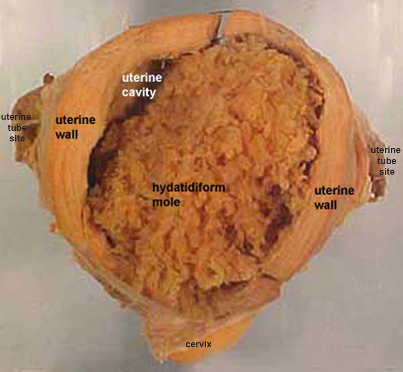
Hydatidiform mole
Several forms of hydatidiform mole: partial mole, complete mole and persistent gestational trophoblastic tumor. (More? mole types). Many of these tumours arise from a haploid sperm fertilizing an egg without a female pronucleus (the alternative form, an embryo without sperm contribution, is called parthenogenesis). The tumour has a "grape-like" placental appearance without enclosed embryo formation. Following a first molar pregnancy, there is approximately a 1% risk of a second molar pregnancy.
ACT Pathology Description
- Macroscopic: The specimen is an enlarged uterus (140x140x100mm, weighing 725g) with right and left ovaries and fallopian tubes attached. Opening the uterus shows a cavity filled with a grape-like mass. The wall of the uterus is thickened and fibrous.
- Microscopic: The sections show an interlacing structure within the fibrous lesion. Sections of the cystic contents show numerous dilated and mucoid degenerate proliferative chorionic villi.
Image Source: ACT Pathology, used with permission.
(More? Week 2 Abnormalities - Hydatidiform Mole ACT Pathology - Uterus benign fibromyoma hydatidiform mole)
File history
Click on a date/time to view the file as it appeared at that time.
| Date/Time | Thumbnail | Dimensions | User | Comment | |
|---|---|---|---|---|---|
| current | 21:38, 8 October 2015 | | 800 × 739 (73 KB) | Z8600021 (talk | contribs) | |
| 11:02, 18 August 2009 |  | 400 × 369 (29 KB) | MarkHill (talk | contribs) | Hydatidiform mole Several forms of hydatidiform mole: partial mole, complete mole and persistent gestational trophoblastic tumor. (More? mole types). Many of these tumours arise from a haploid sperm fertilizing an egg without a female pronucleus (the alt |
You cannot overwrite this file.
File usage
The following 23 pages use this file:
- 2009 Lecture 8
- 2010 BGD Practical 3 - Week 2 Summary
- 2010 BGD Tutorial - Applied Embryology and Teratology
- 2010 Lecture 8
- 2011 Lab 2 - Week 2
- ANAT2341 Lab 2 - Week 2
- ANAT2341 Lab 4 - Abnormal Placenta
- ASA Meeting 2013 - Placenta
- Abnormal Development - Hydatidiform Mole
- BGDA Practical 3 - Week 2 Summary
- BGDA Practical Placenta - Abnormalities
- BGD Tutorial - Applied Embryology and Teratology
- Implantation
- Lecture - Fertilization
- Lecture - Placenta Development
- P
- Placenta - Abnormalities
- Placenta Development
- Trophoblast
- Week 2
- Talk:2011 Lab 2 - Week 2
- File:Placenta abnormalities.jpg
- Template:Placenta Abnormalities gallery