File:Human embryo head week 6 to 8.jpg
Original file (540 × 780 pixels, file size: 66 KB, MIME type: image/jpeg)
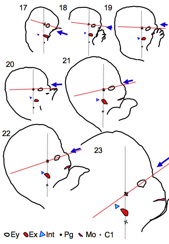
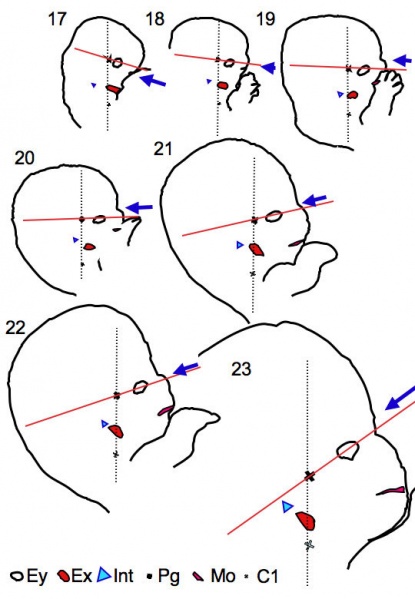
Lateral view of Embryos between Week 6 to 8 (Carnegie Stage 17 to 23) showing Craniofacial Morphogenesis
The dashed line indicates the reference axis (Z-axis) in Method-1 and the red line indicates the reference axis (X-axis) in Method-2.
The arrow indicates the frontal side of the face defined in Method-2.
The following anatomical landmarks are drawn: external ear (Ex), internal ear (int), eye (Ey), mouth (Mo), pituitary gland (Pg), and first cervical vertebra (C1).
Magnification is identical in all stages. The frontal position defined by Method-2 rotated as stages proceed. Note that both external and internal ears were located at similar positions between Pg and C1 between CS 17 and 23
Reference
<pubmed>22296782</pubmed>| PMC3286420 | Head Face Med.
© 2012 Kagurasho et al; licensee BioMed Central Ltd.
This is an Open Access article distributed under the terms of the Creative Commons Attribution License (http://creativecommons.org/licenses/by/2.0), which permits unrestricted use, distribution, and reproduction in any medium, provided the original work is properly cited.
Original file name: Figure 7. 1746-160X-8-2-7.jpg
File history
Click on a date/time to view the file as it appeared at that time.
| Date/Time | Thumbnail | Dimensions | User | Comment | |
|---|---|---|---|---|---|
| current | 22:53, 16 May 2012 | | 540 × 780 (66 KB) | Z8600021 (talk | contribs) | ==Lateral view of embryos between CS 17 and CS 23 showing craniofacial morphogenesis== Human embryo head week 6 to 8.jpg The dashed line indicates the reference axis (Z-axis) in Method-1 and the red line indicates the reference axis (X-axis) in Method-2 |
You cannot overwrite this file.
File usage
The following 3 pages use this file: