File:Human brain growth 01.jpg
From Embryology
Size of this preview: 766 × 600 pixels. Other resolution: 1,022 × 800 pixels.
Original file (1,022 × 800 pixels, file size: 119 KB, MIME type: image/jpeg)
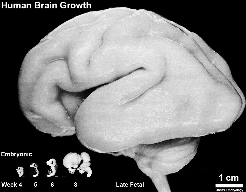
Human Brain Growth
Left lateral view of the relative overall size of the developing human brain during the embryonic period (week 4, 5, 6, and 8) and late fetal (third trimester close to term). Note also the relative size of the spinal cord seen at the lower end of each brain.
| Embryonic Central Nervous System | |||
|---|---|---|---|
| Stage 13 | Stage 14 | Stage 16 | Stage 21 |

scale bar = 1 mm |

|

|

|
| Week 4 | Week 5 | Week 6 | Week 8 |
- Human CNS Images: Carnegie stage 13 | Carnegie stage 13 label | Carnegie stage 14 | Carnegie stage 14 label | Carnegie stage 16 | Carnegie stage 16 label | CN V | Carnegie stage 21 lateral | Carnegie stage 21 median | Fetus CRL 240mm | Neural System Development | Cranial Nerves
Cite this page: Hill, M.A. (2024, April 27) Embryology Human brain growth 01.jpg. Retrieved from https://embryology.med.unsw.edu.au/embryology/index.php/File:Human_brain_growth_01.jpg
- © Dr Mark Hill 2024, UNSW Embryology ISBN: 978 0 7334 2609 4 - UNSW CRICOS Provider Code No. 00098G
File history
Click on a date/time to view the file as it appeared at that time.
| Date/Time | Thumbnail | Dimensions | User | Comment | |
|---|---|---|---|---|---|
| current | 15:11, 7 February 2016 | | 1,022 × 800 (119 KB) | Z8600021 (talk | contribs) |
You cannot overwrite this file.
File usage
The following 6 pages use this file: