File:Huber1915 1fig01.jpg
Huber1915_1fig01.jpg (679 × 600 pixels, file size: 61 KB, MIME type: image/jpeg)
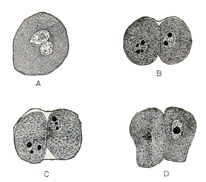
Fig. 1 Tubal ova albino rat
Fig. 1 Tubal ova, albino rat. X 200. A, rat No. 110, 24 hours, 15 min., ovum in pronuclear stage, larger nucleus female pronucleus; B, and C, rat No. 59, 2 days, 2-cell stages, thin oolemma showing in C, only partially seen in B; D, rat No. 62, 2 days, 22 hours, 3-cell stage, the nucleus of the unsegmented blastomere in the monaster phase, only one of the other two cells showing in the figure.
| Historic Disclaimer - information about historic embryology pages |
|---|
 |
- Albino Rat Links: Fig 14. Right Oviduct | Fig 15. 8 and 11-cell stages | The Development of the Albino Rat 1915
Cite this page: Hill, M.A. (2024, May 21) Embryology Huber1915 1fig01.jpg. Retrieved from https://embryology.med.unsw.edu.au/embryology/index.php/File:Huber1915_1fig01.jpg
- © Dr Mark Hill 2024, UNSW Embryology ISBN: 978 0 7334 2609 4 - UNSW CRICOS Provider Code No. 00098G
| Historic Disclaimer - information about historic embryology pages |
|---|
|
Cite this page: Hill, M.A. (2024, May 21) Embryology Huber1915 1fig01.jpg. Retrieved from https://embryology.med.unsw.edu.au/embryology/index.php/File:Huber1915_1fig01.jpg
- © Dr Mark Hill 2024, UNSW Embryology ISBN: 978 0 7334 2609 4 - UNSW CRICOS Provider Code No. 00098G
File history
Click on a date/time to view the file as it appeared at that time.
| Date/Time | Thumbnail | Dimensions | User | Comment | |
|---|---|---|---|---|---|
| current | 10:27, 7 April 2013 | | 679 × 600 (61 KB) | Z8600021 (talk | contribs) | |
| 00:22, 5 April 2013 | 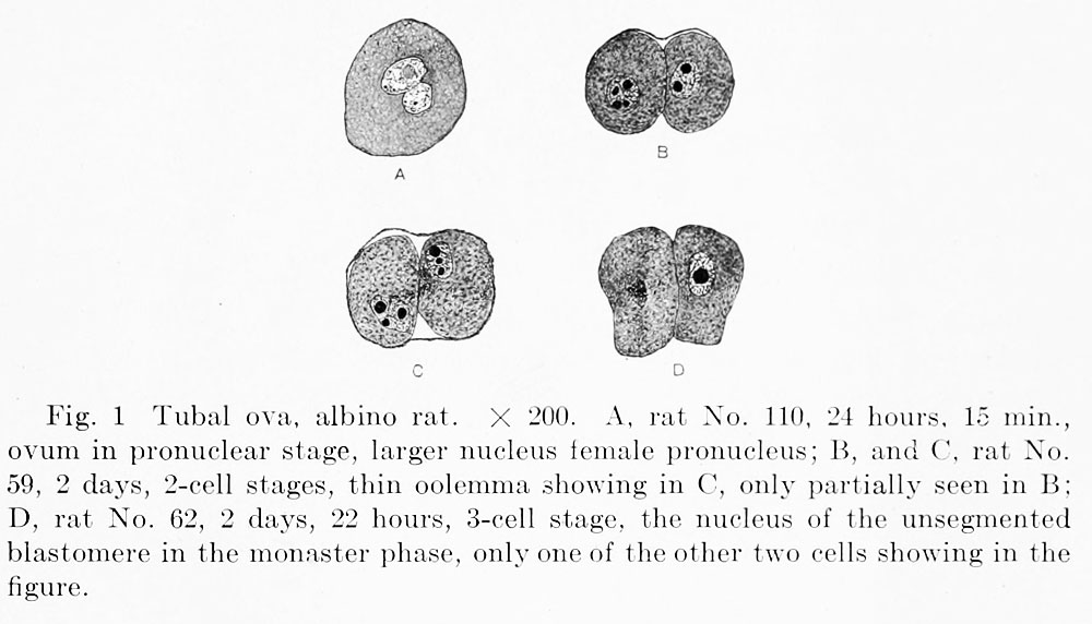 | 1,000 × 572 (79 KB) | Z8600021 (talk | contribs) | {{Huber1915 figures}} {{Huber1915_footer}} |
You cannot overwrite this file.
File usage
The following 2 pages use this file: