File:Heart histology 003.jpg
Heart_histology_003.jpg (400 × 500 pixels, file size: 136 KB, MIME type: image/jpeg)
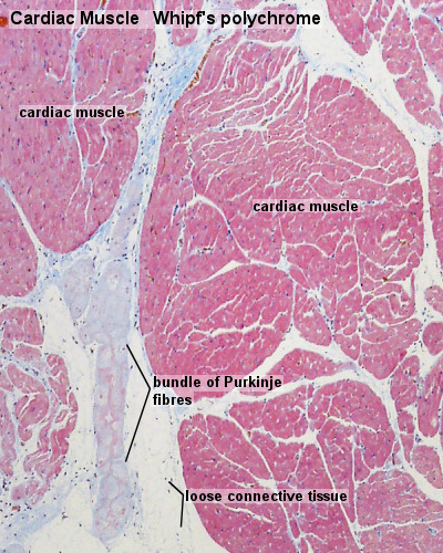
Heart Histology
cardiac muscle, Purkinje Fibre, sheep - Whipf's polychrome transverse section, x40
Whipf's polychrome staining procedure - used by Dr. Louise Whipf, Veterinary Pathology Laboratory, University of Wisconsin, who originally modified Shorr’s (1941) stain.
Original file name: Cardm004wp.jpg
Links: Histology | Histology Stains | Blue Histology images copyright Lutz Slomianka 1998-2009. The literary and artistic works on the original Blue Histology website may be reproduced, adapted, published and distributed for non-commercial purposes. See also the page Histology Stains.
Cite this page: Hill, M.A. (2024, May 21) Embryology Heart histology 003.jpg. Retrieved from https://embryology.med.unsw.edu.au/embryology/index.php/File:Heart_histology_003.jpg
- © Dr Mark Hill 2024, UNSW Embryology ISBN: 978 0 7334 2609 4 - UNSW CRICOS Provider Code No. 00098G
Cite this page: Hill, M.A. (2024, May 21) Embryology Heart histology 003.jpg. Retrieved from https://embryology.med.unsw.edu.au/embryology/index.php/File:Heart_histology_003.jpg
- © Dr Mark Hill 2024, UNSW Embryology ISBN: 978 0 7334 2609 4 - UNSW CRICOS Provider Code No. 00098G
File history
Click on a date/time to view the file as it appeared at that time.
| Date/Time | Thumbnail | Dimensions | User | Comment | |
|---|---|---|---|---|---|
| current | 09:26, 14 August 2011 | | 400 × 500 (136 KB) | S8600021 (talk | contribs) | Heart histology 001.jpg Cardm004wp.jpg |
You cannot overwrite this file.