File:Gray1178.jpg
Gray1178.jpg (600 × 443 pixels, file size: 47 KB, MIME type: image/jpeg)
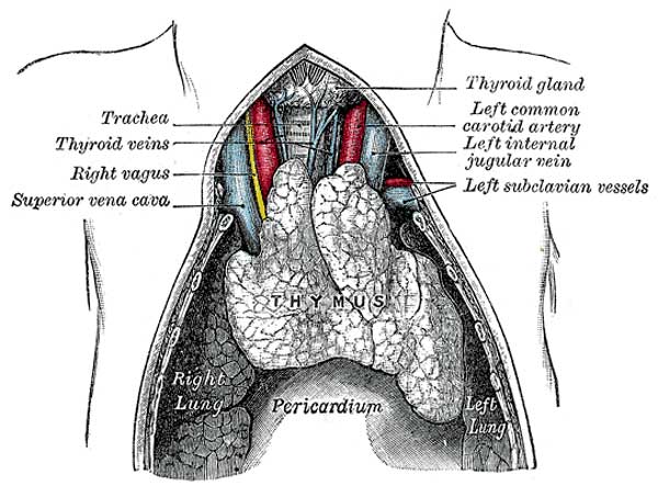
Fig. 1178. The thymus of a full-time fetus
(exposed in situ)
The thymus (Fig. 1178) is a temporary organ, attaining its largest size at the time of puberty (Hammar), when it ceases to grow, gradually dwindles, and almost disappears. If examined when its growth is most active, it will be found to consist of two lateral lobes placed in close contact along the middle line, situated partly in the thorax, partly in the neck, and extending from the fourth costal cartilage upward, as high as the lower border of the thyroid gland. It is covered by the sternum, and by the origins of the Sternohyoidei and Sternothyreoidei. Below, it rests upon the pericardium, being separated from the aortic arch and great vessels by a layer of fascia. In the neck it lies on the front and sides of the trachea, behind the Sternohyoidei and Sternothyreoidei. The two lobes generally differ in size; they are occasionally united, so as to form a single mass; and sometimes separated by an intermediate lobe. The thymus is of a pinkish-gray color, soft, and lobulated on its surfaces. It is about 5 cm. in length, 4 cm. in breadth below, and about 6 mm. in thickness. At birth it weighs about 15 grams, at puberty it weighs about 35 grams; after this it gradually decreases to 25 grams at twentyfive years, less than 15 grams at sixty, and about 6 grams at seventy years.
Cite this page: Hill, M.A. (2024, April 27) Embryology Gray1178.jpg. Retrieved from https://embryology.med.unsw.edu.au/embryology/index.php/File:Gray1178.jpg
- © Dr Mark Hill 2024, UNSW Embryology ISBN: 978 0 7334 2609 4 - UNSW CRICOS Provider Code No. 00098G
File history
Click on a date/time to view the file as it appeared at that time.
| Date/Time | Thumbnail | Dimensions | User | Comment | |
|---|---|---|---|---|---|
| current | 11:09, 24 December 2010 |  | 600 × 443 (47 KB) | S8600021 (talk | contribs) |
You cannot overwrite this file.
File usage
The following 4 pages use this file:
